Case Author(s): Yonglin Pu, M.D.,Ph.D. Jerold Wallis, M.D. , 03/23/03 . Rating: #D3, #Q4
Diagnosis: Extra-Skeletal Ewing's Sarcoma
Brief history:
A 25-year-old man presents with right leg pain and right thigh mass.
Images:

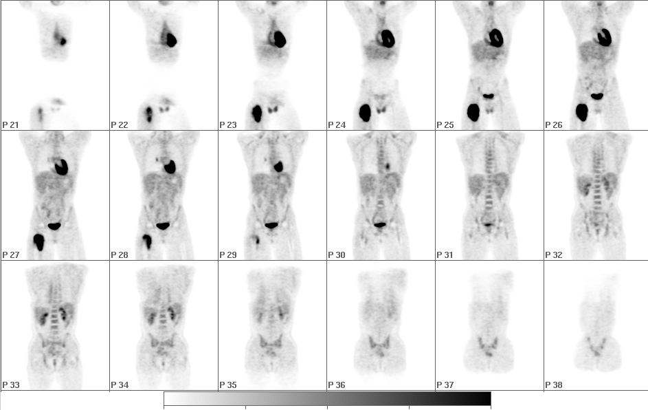
Tumor PET Imaging (Metabolism), Initial staging
View main image(pt) in a separate image viewer
View second image(mr).
MRI of both thighs, T1WI and T2WI
View third image(ct).
CT of Chest with contrast
Full history/Diagnosis is available below
Diagnosis: Extra-Skeletal Ewing's Sarcoma
Full history:
A 25-year-old man initially presented with right leg pain one and a half months ago and was found to have a right thigh mass that was biopsied. Imaging and pathology suggested a diagnosis of Ewing´s sarcoma of an extra-skeletal origin. The patient has not received chemotherapy or radiation therapy at this time and presents for evaluation and staging.
Radiopharmaceutical:
15.0 mCi F-18 Fluorodeoxyglucose
Findings:
There is intensely increased uptake of FDG in a large mass in the right thigh corresponding to the mass seen on the MRI. There is no central photopenia identified and the activity appears relatively homogeneous. Also, there is a focus of mildly intense uptake in the high right inguinal region (frame 25), which could represent metastasis or inflammation. There is a focus of moderately increased uptake in the right hilum of the lung (frame 28), suspicious for metastasis. Normal muscle activity is noted in the right upper extremity at the insertion of the deltoid muscle at the right humerus and in the lower neck.
Discussion:
Extraosseous Ewing's Sarcoma is a rare soft tissue sarcoma. It is composed of small undifferentiated round to oval cells. It is histologically and ultrastructurally indistinguishable from the osseous form. The diagnosis of Extraosseous Ewing's Sarcoma is made on the basis of histological findings in the absence of bony involvement at the time of presentation.There are only a few case reports about MRI findings of Extraosseous Ewing's Sarcoma.
According to our limited literature search from Medline and Premedline from 1966 to 2003, there are no reports about PET findings of Extraosseous Ewing's Sarcoma. Our case showed that it has intense and homogenous FDG uptake in a large tumor mass in the soft tissue of right thigh with multiple sites of metastases. The metastatic lesions also showed intense FDG uptake.
References:
1. Ming L, Reu S, Chiao Y, Gin C Extraosseous Ewing's Sarcoma: A Case Report with MR Manifestations. Chin J Radiol 2000; 25: 251.
2. Mukhopadhyay P. Gairola M. Sharma M. Thulkar S. Julka P. Rath G. Primary spinal epidural extraosseous Ewing's sarcoma: report of five cases and literature review. Australasian Radiology. 2001;45(3):372.
3. Thebert A. Francis IR. Bowerman RA. Retroperitoneal extraosseous Ewing's sarcoma with renal involvement: US and MRI findings. Clinical Imaging. 1993; 17(2):149.
Followup:
The patient has been through six cycles of chemotherapy. The previously described intense uptake of FDG in the large mass in the right thigh has markedly decreased in size and intensity. A heterogeneous area of mild to moderately increased uptake in this region is seen, which is suspicious for residual tumor. A small focus of moderately increased uptake in the right inguinal region noted on the prior study is no longer present on today´s examination. However, the focus of moderately increased uptake in the right hilum has markedly increased in size and intensity, which correlates with the CT finding of an interval increase in size of a right hilar calcified mass and likely represents interval worsening of metastatic disease.
The diagnosis was confirmed by tissue biopsy from mass in the right thigh.
View followup image(pt).
Tumor PET Imaging (Metabolism), restaging
Major teaching point(s):
Extraosseous Ewing's Sarcoma is a rare tumor. This case showed that it has intense and homogenous FDG uptake in the primary and metastatic lesions.
Differential Diagnosis List
1. Liposarcoma 2.Malignant fibrous histiocytoma,3.Rhabdomyosarcoma
ACR Codes and Keywords:
References and General Discussion of PET Tumor Imaging Studies (Anatomic field:Skeletal System, Category:Neoplasm, Neoplastic-like condition)
Search for similar cases.
Edit this case
Add comments about this case
Return to the Teaching File home page.
Case number: pt098
Copyright by Wash U MO

