Case Author(s): Yonglin Pu, M.D., Ph.D. and Jerold Wallis, M.D. , 03/22/2003 . Rating: #D3, #Q4
Diagnosis: Metastatic Breast carcinoma
Brief history:
48 year old woman with a history of breast caner.
Images:
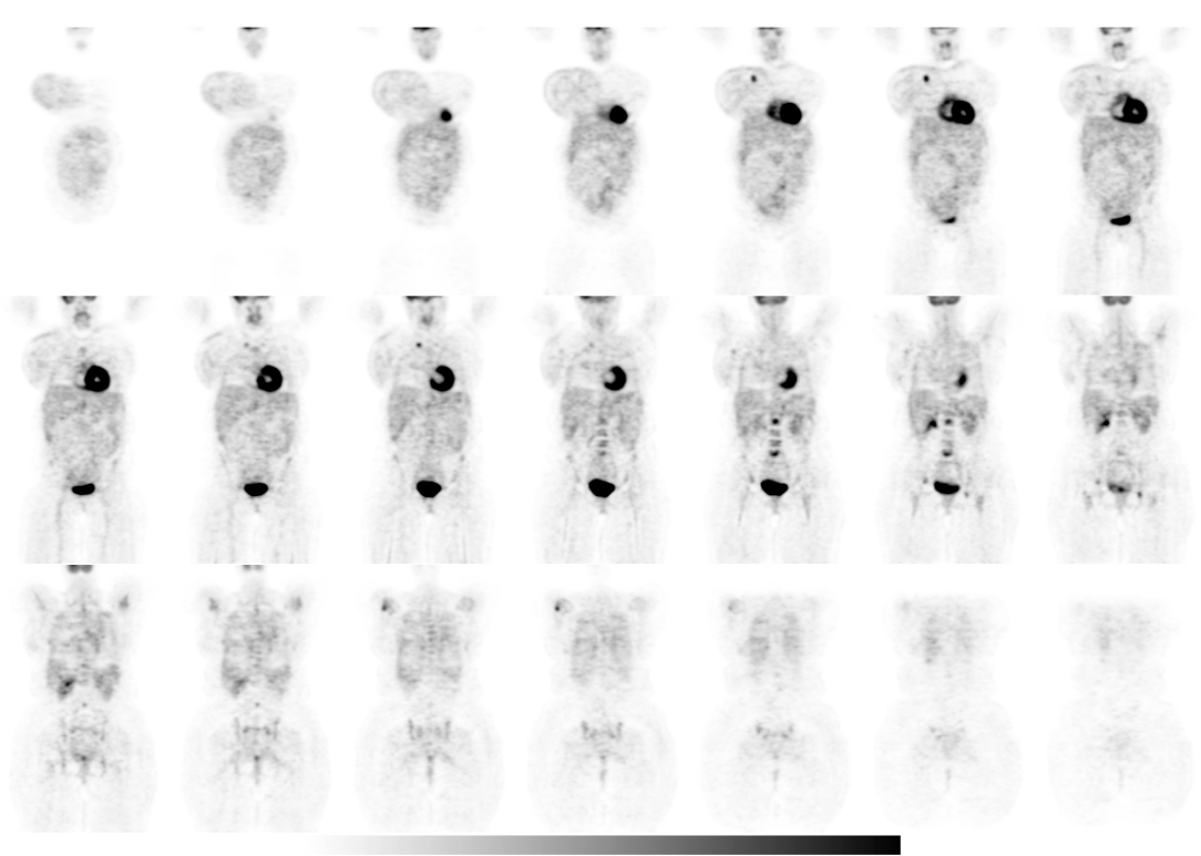
Tumor PET Imaging, Coronal sections
View main image(pt) in a separate image viewer
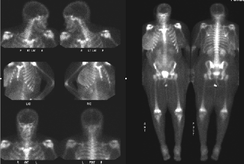
View third image(bs).
Bone Scintigraphy (whole body)
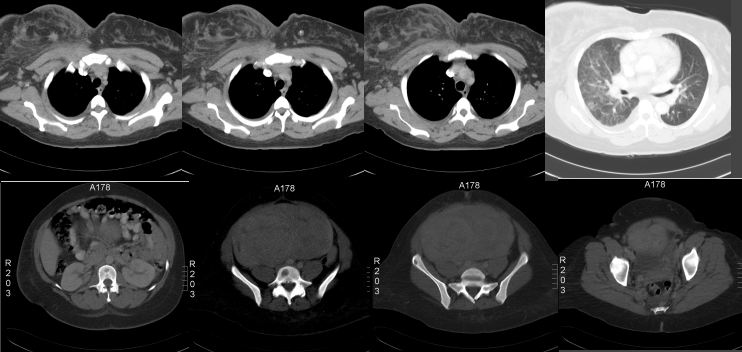
View fourth image(ct).
CT of chest, abdomen and pelvis
Full history/Diagnosis is available below
Diagnosis: Metastatic Breast carcinoma
Full history:
A 48-year-old woman with a history of breast cancer, which was diagnosed by biopsy about a month ago. She started chemotherapy 5 days ago. This tumor PET imaging was performed for initial staging.
Radiopharmaceutical:
15.0 mCi Fluorodeoxyglucose i.v.
Findings:
There is focally increased activity in the upper portion of the right breast, which correlates with the spiculated lesion on the comparison CT images. This finding represents primary breast carcinoma. There is also focally increased activity in the right upper chest wall, approximately at the level of the breast lesion, but medial and deep to the breast lesion, which could represent a regional lymph node or a rib metastasis. There is also focal mildly increased FDG uptake at the tail of the right breast or right axilla which likely represents lymphadenopathy. There is diffuse uptake in the right breast which likely represents inflammatory reaction. Diffuse uptake of a mild degree also is noted in the lower abdomen, which correlates with the large leiomyoma in the uterus on the comparison CT images. There are foci of increased FDG uptake in the L2 and L5 vertebral bodies, the L3 spinous process, the left acetabular region, the right supraacetabular region, and the right humeral head. These findings likely represents osseous metastases. The L2 and L5 lesions correspond to lytic vertebral lesions on the comparison CT images.
A whole body bone scintigraphy performed 33 days ago demonstrated no significant evidence for bony metastasis.
Discussion:
FDG-PET has a better specificity for malignant lesions when compared to bone scintigraphy. Depending on tumor type, some bone metastasis may be better seen on PET FDG imaging, and some better seen on bone scintigraphy. In this case, the lytical lesions demonstrated on CT were clearly seen on FDG PET, but not seen on bone scintigraphy.
Reference:
Kao CH. Hsieh JF. Tsai SC. Ho YJ. Yen RF.Comparison and discrepancy of 18F-2-deoxyglucose positron emission tomography and Tc-99m MDP bone scan to detect bone metastases. Anticancer Research. 20(3B):2189-92, 2000.
Followup:
Needle biopsy showed invasive ductal carcinoma in the right breast.
ACR Codes and Keywords:
References and General Discussion of PET Tumor Imaging Studies (Anatomic field:Breast, Category:Neoplasm, Neoplastic-like condition)
Search for similar cases.
Edit this case
Add comments about this case
Return to the Teaching File home page.
Case number: pt097
Copyright by Wash U MO