Case Author(s): Brock G. McDaniel, M.D. and Farrokh Dehdashti, M.D. , . Rating: #D., #Q.
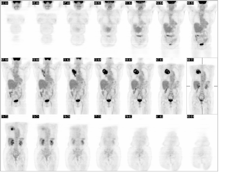
Diagnosis: Non-small cell lung cancer of the right upper lobe with pulmonary vein extension.
Brief history:
68-year-old female with right upper lobe mass.
Images:

Coronal PET
View main image(pt) in a separate image viewer
View second image(xr).
PA chest radiograph
View third image(pt).
PET/CT transaxial
View fourth image(pt).
PET/CT coronal
Full history/Diagnosis is available below
Diagnosis: Non-small cell lung cancer of the right upper lobe with pulmonary vein extension.
Full history:
68-year-old female, with a remote history of tobacco use who presented, in November of 2002, with an acute episode of shortness of breath and cough. A chest x-ray demonstrated a right upper lobe mass with associated pneumonia. A chest CT confirmed a 5cm right upper lobe mass. Also, noted was a 1.1 cm right hilar node. The patient was staged utilizing CT, PET, bronchoscopy and mediastinoscopy.
Radiopharmaceutical:
15 mCi F-18 Fluorodeoxyglucose i.v.
Findings:
The PET/CT images demonstrate intense FDG uptake within the right upper lobe mass, which extends centrally to involve the right hilum. No distant metastasis seen. The chest radiograph (PA) shows a large right upper lobe mass concerning for a primary lung neoplasm. The contrasted CT confirms the right upper lobe mass which measures 8.0 cm x 5.2 cm and also demonstrates right hilar adenopathy (1.1 cm).
Discussion:
Staging of non-small cell lung cancer (NSCLC) is important both for providing prognostic information and guiding selection of treatment strategy. A recent meta-analysis of the literature demonstrated that FDG-PET is more sensitive and specific than CT in evaluating mediastinum. A negative PET provides greater than 90% certainty that the mediastinal lymph nodes are not involved. PET is considered as the most accurate noninvasive method to characterize mediastinal lymph node status. It has been shown that FDG-PET improves patient management in 15-30% of patients.
Reference:
Toloza et al., Chest 2003; 123: 137-146.
Followup:
Per the operative report, during the right upper lobe resection, it became apparent that the tumor extended centrally to involve the right upper and middle lobe veins. Therefor, both the right middle and upper lobes were resected as well as ribs 3, 4 and 5 which had direct invasion from the primary mass. In retrospect, the 1.1-cm right hilar soft tissue mass, originally described as a hilar node, represents tumor thrombus within the superior pulmonary vein. The curvilinear fucus of increased FDG uptake, which extends to the right hilum, also represents the tumor thrombus.
Closer inspection of the contrasted chest CT demonstrates the tumor filled superior pulmonary vein, where one would expect it to be, just anterior to the right pulmonary artery.
View followup image(ct).
Contrasted CT of the chest
Differential Diagnosis List
The differential diagnosis for the curvilinear FDG uptake includes primary extension along the pleura, atelectatic post obstructive pneumonitis, hilar adenopathy and as in this case pulmonary vein extension.
ACR Codes and Keywords:
References and General Discussion of PET Tumor Imaging Studies (Anatomic field:Lung, Mediastinum, and Pleura, Category:Neoplasm, Neoplastic-like condition)
Search for similar cases.
Edit this case
Add comments about this case
Return to the Teaching File home page.
Case number: pt096
Copyright by Wash U MO

