Case Author(s): Brock G. McDaniel, M.D. and Tom R. Miller, M.D., Ph.D. , 05/29/2002 . Rating: #D3, #Q3
Diagnosis: Patient Motion Artifact
Brief history:
54 year old female with left upper lobe mass.
Images:

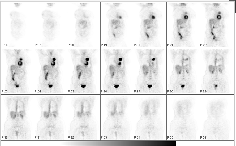
Coronal PET images
View main image(pt) in a separate image viewer
Full history/Diagnosis is available below
Diagnosis: Patient Motion Artifact
Full history:
54 year old female smoker with a routine chest x-ray and follow-up chest CT which demonstrated a large left upper lobe mass and several small pleural-based nodules.
Findings:
There is intense FDG uptake in the left upper lobe mass consistent with malignancy; no metastasis are seen.
There is a peripheral band of apparent increased activity along the left lateral and posterior hemithorax. Also, there is more subtly increased activity along the contour of the right body surface.
Discussion:
Patient motion between the transmission and emission scans can lead to artifacts as seen above. Movement prior to completion of the study will cause the attenuation map obtained from the transmission scan to be incorrectly aligned with the emission data, thereby creating regions of under-correction and over-correction. This is readily evident in this patient where we see a band of apparent increased activity along the left pleura. This is also apparent more subtly along the right body surface.
Followup:
The follow-up scan performed approximately 4 month later demonstrates significant improvement in scan quality with minimal motion artifact. However, the patient apparently shifted slightly in the opposite direction because there now is a subtle band of increased activity along the right pleura.
View followup image(pt).
Repeat whole body PET scan approximately 4 months later
Differential Diagnosis List
In the appropriate clinical setting and without evidence of motion, post radiation changes involving the pleura of the left hemithorax would be a consideration. Conceivably, a malignant pleural effusion might also have a similar appearance.
ACR Codes and Keywords:
References and General Discussion of PET Tumor Imaging Studies (Anatomic field:Lung, Mediastinum, and Pleura, Category:Other(Artifact))
Search for similar cases.
Edit this case
Add comments about this case
Return to the Teaching File home page.
Case number: pt095
Copyright by Wash U MO

