Case Author(s): Christian T. Schmitt, M.D. and Tom R. Miller, M.D., Ph.D. , 1/15/03 . Rating: #D3, #Q4
Diagnosis: Adenocarcinoma of the sigmoid colon and hepatic metastases in a lung transplant patient
Brief history:
36 year old man with a history of cystic fibrosis who underwent bilateral lung transplantation 9 years ago, now presenting with an upper abdominal mass.
Images:
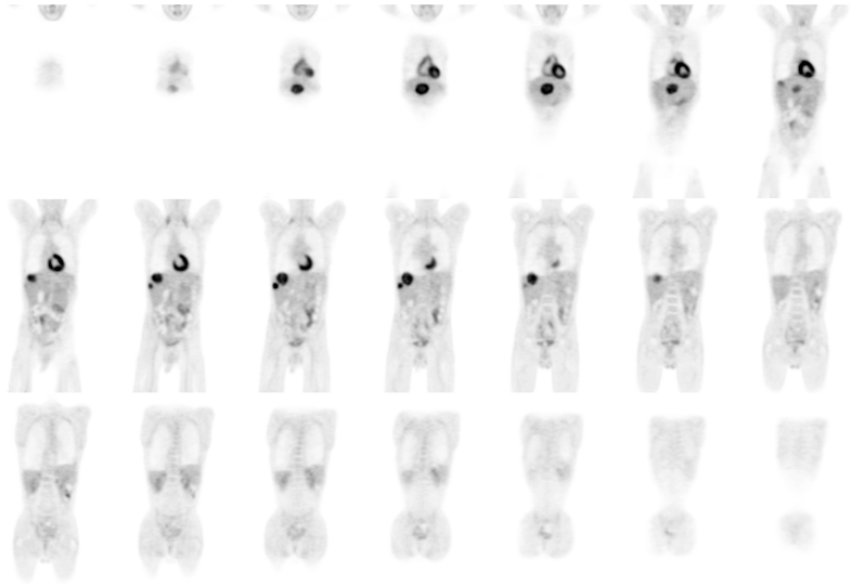
Coronal whole body images
View main image(pt) in a separate image viewer
View second image(pt).
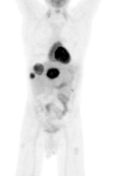
Rotating projection images
View third image(ct).
Selected axial contrast-enhanced CT images
View fourth image(xr).
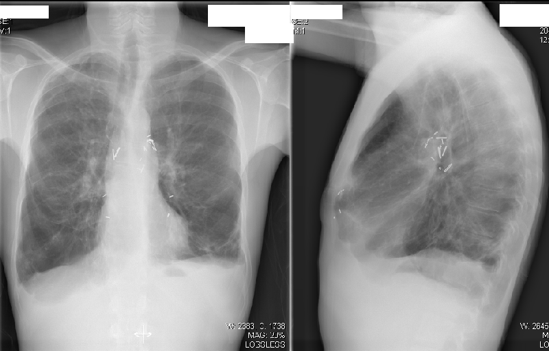
Chest radiograph
Full history/Diagnosis is available below
Diagnosis: Adenocarcinoma of the sigmoid colon and hepatic metastases in a lung transplant patient
Full history:
The patient is a 36 year old man with a history of cystic fibrosis who underwent bilateral lung transplantation 9 years ago. He recently presented with an upper abdominal mass. Subsequent evaluation revealed adenocarcinoma of the sigmoid colon with hepatic metastases. The patient presented for PET imaging for additional staging evaluation.
Radiopharmaceutical:
14.9 mCi F-18 Fluorodeoxyglucose I.V.
Findings:
PET Study: 3 large lesions within the liver demonstrate marked uptake of FDG and correspond to the lesions on CT and are compatible with hepatic metastases. No other areas of abnormal uptake are noted.
CT Study: 3 large low-density lesions with some enhancement compatible with hepatic metastases.
Chest Radiograph: Post-surgical changes compatible with the history of bilateral lung transplantation.
Discussion:
This case demonstrates one the the possible complications of lung transplantation, the development of colon cancer. Abdominal complications from lung transplantation can range from infectious or inflammatory conditions, such as diverticulitis, CMV colitis or toxic megacolon, to neoplastic conditions like polyps, lymphoproliferative disorder or cancer.
The PET study in this case confirmed the known hepatic metastases and did not demonstrate additional disease. PET has been shown to be very useful in the staging and restaging of colorectal carcinoma. PET can also be useful in the evaluation of post-transplant lymphoproliferative disorder.
ACR Codes and Keywords:
- General ACR code: 73
- Gastrointestinal System:
7.33 "MALIGNANT NEOPLASM-SECONDARY (METASTATIC)"
References and General Discussion of PET Tumor Imaging Studies (Anatomic field:Gasterointestinal System, Category:Neoplasm, Neoplastic-like condition)
Search for similar cases.
Edit this case
Add comments about this case
Return to the Teaching File home page.
Case number: pt093
Copyright by Wash U MO