Case Author(s): Brock McDaniel, M.D. and Tom R. Miller, M.D., Ph.D. , 9/05/02 . Rating: #D3, #Q4
Diagnosis: Endometrial cancer with Sister Mary Joseph nodule
Brief history:
55 year old woman with history of endometrial cancer.
Images:

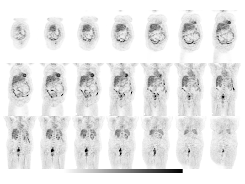
Coronal FDG-PET
View main image(pt) in a separate image viewer
View second image(pt).
Transaxial FDG-PET
View third image(ct).
Abdominal CT
View fourth image(ct).
Pelvic CT
Full history/Diagnosis is available below
Diagnosis: Endometrial cancer with Sister Mary Joseph nodule
Full history:
This 56 year old woman has a history of clinical stage II endometrial cancer. She is being evaluated for possible radiation therapy and/or chemotherapy.
Radiopharmaceutical:
15 mCi F-18 Fluorodeoxyglucose i.v.
Findings:
The PET images demonstrate marked uptake within the cervix and uterus consistent with the history of Stage II endometrial carcinoma. Additional activity is noted in a calcified mass adjacent to the anterior aspect of the uterine fundus, seen on the CT image through the pelvis. This was felt on initial interpretation of the CT to represent infiltration of the endometrial carcinoma into a uterine leiomyoma. Spread to an adjacent right ovary was also entertained, but believed to be less likely. A focus of intense FDG uptake is seen in the peri-umbilical region (especially transaxial slices p55 and p56). The CT images demonstrate a peri-umbilical nodule corresponding to the focus of increased PET uptake.
Discussion:
In this patient who was original felt to have clinical Stage II endometrial cancer, PET imaging was able to demonstrate distant metastasis to a peri-umbilical lymph node, referred to as a Sister Mary Joseph Node. The identification of this nodule up-staged the patient to clinical Stage 4B. In general, a Sister Mary Joseph node is associated with advanced disease and signifies poor prognosis. Other primary neoplasms associated with these peri-umbilical nodules include gastric, ovarian, colorectal and pancreatic carcinomas.
Historically, Sister Mary Joseph was a surgical assistant under the guidance of Dr. William Mayo. While performing her duties preparing the patientsí abdomen for surgery, she identified the peri-umbilical nodule, which would come to bear her name. It was noted that the presence of this nodule often indicated advanced disease.
Major teaching point(s):
The presence of a Sister Mary Joseph Nodule/Node implies a poor prognosis.
Differential Diagnosis List
Peri-umbilical tumors can be either benign or malignant. Benign tumors make up about 57% of all umbilical tumors. Such benign causes include dermal nevi, fibroepithelial papillomas and dermatofibromas to name a few. Malignant causes make up about 43%. Primary tumors are exceedingly rare and include adenocarcinomas and myosarcomas arising from remnants of the omphalomesenteric duct or urachus. Metastatic tumors make up 83% of malignant periumbilical tumors and includes endometrial, gastric, ovarian, colorectal and pancreatic carcinomas.
ACR Codes and Keywords:
References and General Discussion of PET Tumor Imaging Studies (Anatomic field:Genitourinary System, Category:Neoplasm, Neoplastic-like condition)
Search for similar cases.
Edit this case
Add comments about this case
Return to the Teaching File home page.
Case number: pt091
Copyright by Wash U MO

