Case Author(s): Jayson R. St. Jacques, M.D. and Barry A. Siegel, M.D. , 12/2/02 . Rating: #D4, #Q4
Diagnosis: Non-Small Cell Lung Cancer with Transmission Imaging Artifact.
Brief history:
57-year-old woman with history of right lung mass.
Images:

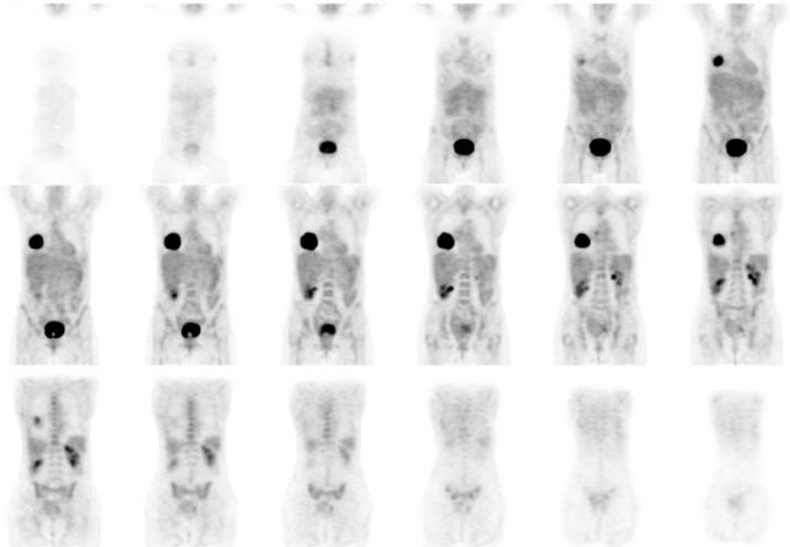
Coronal whole-body FDG-PET images.
View main image(pt) in a separate image viewer
View second image(pt).
Whole-body coronal transmission image with associated attenuation- corrected emission image.
View third image(ct).
Axial bone-window and soft-tissue-window CT images, transmission PET image, and attenuation-corrected PET image at the level of the mass.
View fourth image(mc).
Germanium-68 decay scheme
Full history/Diagnosis is available below
Diagnosis: Non-Small Cell Lung Cancer with Transmission Imaging Artifact.
Full history:
57-year-old woman with a recently diagnosed non-small cell lung cancer. FDG-PET was requested for initial staging.
Radiopharmaceutical:
12.3 mCi F-18 Flurodeoxyglucose i.v.
Findings:
There is a large right lower lobe mass with central necrosis, consistent with the patient's known lung cancer. The maximum standardized uptake value (SUV) of this lesion was 17.4, consistent with malignancy. An additional focus of increased FDG uptake within a subcarinal lymph node is suspicious for metastatic disease.
Discussion:
This case demonstates an artifact associated with post-emission transmission imaging (i.e., transmission imaging that is performed after the patient has been injected with FDG). Post-emission transmission imaging is commonly performed PET scanners that utilize rotating Germanium-68 rod sources for transmission imaging. The transmission imaging data are used for for attenuation correction of the emission images. Although such transmission data sets are always "contaminated" with emission data (unless an explicit correction for this is applied to the transmission data), the effect is usually small. In the case of very "hot" tumors (such as in this patient), the large amount of activity within the lesion leads to an underestimation of the attenuation value of the lesion; in this case, the transmission image shows the majority of the lesion to have an attenuation value similar to that of lung rather than that of soft tissue. Accordingly, the emission image is undercorrected, and the measured SUV in the tumor will be artifactually lower than the true valuse. A similar phenomenon occurs with excreted FDG in the bladder, as seen to a lesser extent on the coronal comparison images (second image set).
Germanium-68, with a half life of 270.8 days, decays by electron capture to Gallium-68, which has a half life of 67.6 minutes, which then decays by electron capture and positron emission to stable Zinc-68. It is the annihilation radiation from the positrons emitted by the decay of Ga-68 that is used for transmission imaging.
Major teaching point(s):
For the reasons stated above, the SUVs of very "hot" structures is likely to be underestimated on PET obtained with post-emission transmission imaging. In the case of the differential diagnosis of lung nodules, this effect is likely to be of little significance, because a lesion having an SUV close to the threshold for diagnosis of malignancy (typically stated to be 2.5) wil make a small contribution to the transmission measurement.
ACR Codes and Keywords:
References and General Discussion of PET Tumor Imaging Studies (Anatomic field:Lung, Mediastinum, and Pleura, Category:Neoplasm, Neoplastic-like condition)
Search for similar cases.
Edit this case
Add comments about this case
Return to the Teaching File home page.
Case number: pt090
Copyright by Wash U MO

