Case Author(s): Yonglin Pu, MD,Ph.D. Farrokh Dehdashti,MD , 03/20/03 . Rating: #D3, #Q3
Diagnosis: Malignant Mesothelioma
Brief history:
62 year old woman with history of remote asbestos exposure presents with left chest discomfort.
Images:
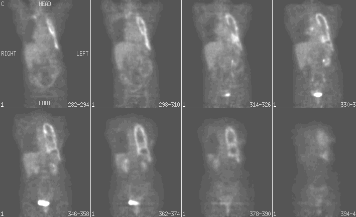
Tumor PET Imaging (Metablism)
View main image(pt) in a separate image viewer
View second image(ct).
Chest CT with contrast
Full history/Diagnosis is available below
Diagnosis: Malignant Mesothelioma
Full history:
62-year-old woman presents with pleural effusion. She has a history of remote asbestos exposure.
Radiopharmaceutical:
2.7 mCi F-18 Fluorodeoxyglucose intravenously
Findings:
Intense nodular uptake is seen involving the pleura of the entire left hemithorax corresponding to the concentric, diffuse and nodular pleural thickening on the comparison CT images. The foci of FDG uptake are seen in a subcarinal lymph node and right anterosuperior mediastinum, corresponding to the lymphadenopathy on the CT images performed three days ago. The extensive volume loss of the left lung is noted. There is no involvement of the right pleura.
Discussion:
The diagnosis of malignant mesothelioma is difficult with CT, which often cannot differentiate between benign pleural thickening and malignant pleural abnormalities such as mesothelioma and pleural metastases. Thoracentesis and CT-guided biopsy are invasive and sometime inconclusive. FDG-PET has been used in the diagnosis of mesothelioma. Bury et al have demonstrated that FDG-PET has a sensitivity of 94% and specificity of 78% for differentiating benign from malignant pleural abnormalities. Similar results have been reported by Lowe et al (sensitivity of 94% and specificity of 67%). A malignant pleural disease typically exhibits moderate to markedly increased FDG accumulation, which often has a nodular pattern. A lack of FDG uptake within the pleural lesion has a high negative predictive value for absence of malignant disease. In both reports, false-positive results were seen in patients with infectious/inflammatory pleural disease. Talc Pleurodesis can also simulate a pleural malignancy. In another study, a standard uptake value (SUV) with a cutoff of 2.0 differentiates between malignant and benign pleural disease with a sensitivity of 91% and a specificity of 100%. A recent study demonstrated that FDG-PET is also useful for the staging of malignant mesothelioma in patients who were candidates for aggressively combined modality therapy (4). FDG-PET identified all 18 primary malignant mesotheliomas with a mean standard uptake value (SUV) of 7. 6. In two patients, extrathoracic metastases in the axillary and cervical lymph node and bone metastases were demonstrated by PET. They were excluded from surgery. Two false positive results were noted in this study, one from contralateral pleural inflammation, and another from post-surgical change in the peritoneal cavity for the treatment of diverticulitis.
References:
1) Knight SB, Delbeke D, Stewart JR, Sandler MP. Evaluation of pulmonary lesions with FDG-PET. Comparison of findings in patients with and without a history of prior malignancy. Chest 1996 Apr; 109(4):982-8.
2) Lowe VJ, Patz E, Harris L, Hoffman JM, Hanson M, Goodman P, Coleman RE. FDG-PET evaluation of pleural abnormalities. J Nucl Med 1994; 35:228P.
3). Benard F, Sterman D, Smith RJ, et al. Metabolic imaging of malignant pleural mesothelioma with fluorodeoxyglucose positron emission tomography. Chest 1998; 114(3): 713-722.
4). Schneider DB. Clary-Macy C. Challa S. Sasse KC. Merrick SH. Hawkins R. Caputo G. Jablons D. Positron emission tomography with F 18-fluorodeoxyglucose in the staging and preoperative evaluation of malignant pleural mesothelioma. Journal of Thoracic & Cardiovascular Surgery. 2000; 120(1):128-33
Followup:
Pleural biopsy demonstrated malignant mesothelioma(epithelial type).
Differential Diagnosis List
Pleural metastases and pleural change from Talc Pleurodesis
ACR Codes and Keywords:
References and General Discussion of PET Tumor Imaging Studies (Anatomic field:Lung, Mediastinum, and Pleura, Category:Neoplasm, Neoplastic-like condition)
Search for similar cases.
Edit this case
Add comments about this case
Return to the Teaching File home page.
Case number: pt088
Copyright by Wash U MO