Case Author(s): Christian T. Schmitt, M.D. and Barry A. Siegel, M.D. , 10/14/02 . Rating: #D3, #Q4
Diagnosis: Sarcoidosis
Brief history:
46-year-old woman with newly diagnosed cervical carcinoma presents for staging
Images:
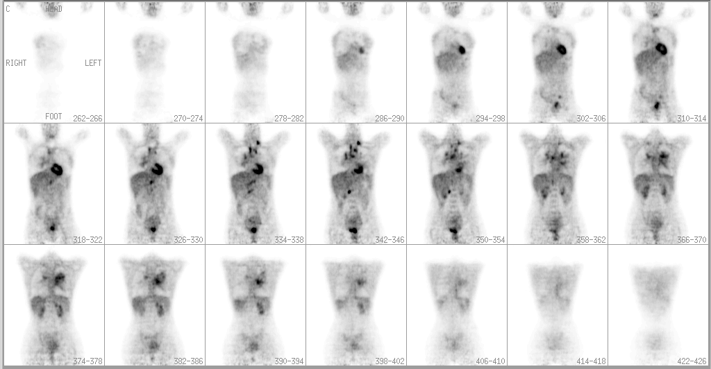
Coronal whole body FDG-PET images are shown
View main image(pt) in a separate image viewer
View second image(ct).
CT images of the chest
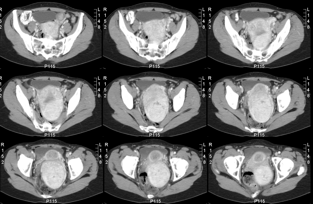
View third image(ct).
CT images of the abdomen and pelvis
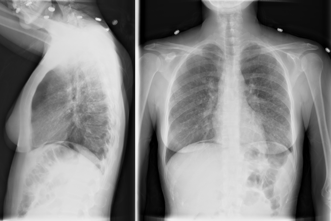
View fourth image(xr).
Chest radiograph
Full history/Diagnosis is available below
Diagnosis: Sarcoidosis
Full history:
46-year-old woman initially presented with vaginal bleeding. She was subsequently diagnosed with cervical adenocarcinoma. She presented for staging of her known carcinoma. There was no other significant past medical history at that time.
Radiopharmaceutical:
2.3 mCi F-18 Fluorodeoxyglucose i.v.
Findings:
8-14-02 PET: There is increased uptake within a left supraclavicular lymph node, as well as hilar and mediastinal lymph nodes. There is also increased lung parenchymal uptake in a perihilar distribution. Additionally there is increased uptake in paraaortic retroperitoneal lymph nodes and left external iliac chain lymph nodes. Increased uptake in the pelvis consistent with the primary cervical carcinoma is seen with extension to the uterine fundus.
8-14-02 CT: Nodular interstitial lung disease with mediastinal lymphadenopathy suggestive of lymphangitic spread of carcinoma. Cervical mass consistent with known primary carcinoma. No abdominal or pelvic lymphadenopathy.
8-29-02 CXR: Bilateral reticulonodular interstitial opacities.
Discussion:
Cervical cancer typically spreads by direct extension and by orderly lymphatic spread first to pelvic, then to para-aortic, and then to supraclavicular modes. Spread by the hematogenous route occurs later. The unusual part of this case was the perihilar and mediastinal involvement in the chest with the appearance more typical of lymphangitic spread of tumor in the chest, rather than mostly peripheral parenchymal involvement as one would expect with hematogenous lung metastases. This prompted further evaluation of the left supraclavicular lymph node.
Followup:
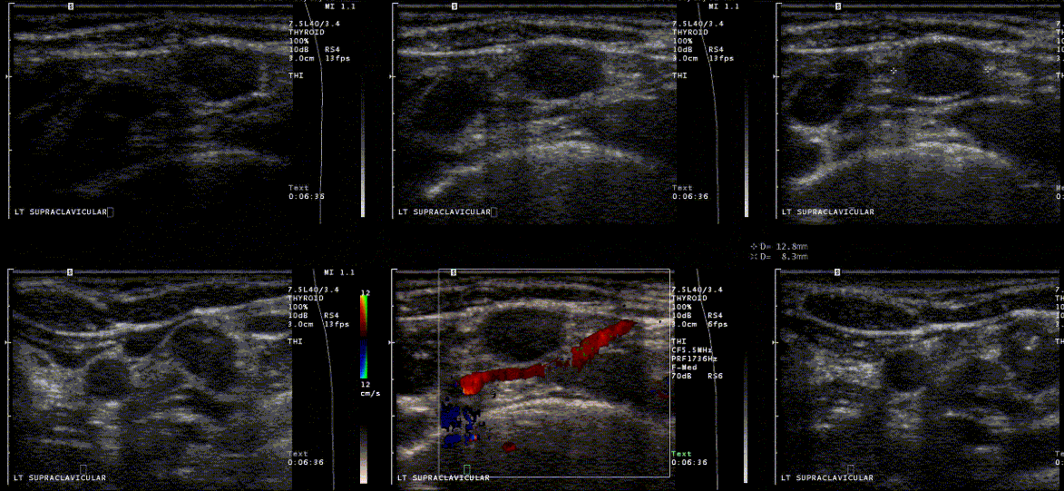
The patient had an ultrasound-guided biopsy of a 1.2 x 0.8 cm left supraclavicular lymph node. (The ultrasound guidance images are provided below.) The pathology demonstrated granulomas and multinucleated giant cells suggestive of sarcoidosis. Subsequent transbronchial biopsy also revealed non-caseating granulomas compatible with sarcoidosis
The patient subsequently underwent exploratory laparotomy with lymph node dissection. The primary cervical adenocarcinoma was confirmed as was metastasis to pelvic, left external iliac, and paraaortic lymph nodes.
View followup image(us).
Major teaching point(s):
This is not the first example of dramatic FDG uptake with sarcoidosis in the teaching file. It is important to keep in mind as a potential cause of a false-positive FDG-PET study. Particularly, it may be helpful to consider sarcoidosis when the pattern of nodal involment is not entirely typical for the primary tumor being evaluated, as in this case.
ACR Codes and Keywords:
- General ACR code: 62
- Lung, Mediastinum, and Pleura:
6.22 "SARCOIDOSIS"
References and General Discussion of PET Tumor Imaging Studies (Anatomic field:Lung, Mediastinum, and Pleura, Category:Inflammation,Infection)
Search for similar cases.
Edit this case
Add comments about this case
Return to the Teaching File home page.
Case number: pt087
Copyright by Wash U MO