Case Author(s): Jayson R. St. Jacques, M.D. and Tom R. Miller, M.D., Ph.D. , 10/18/02 . Rating: #D2, #Q4
Diagnosis: Metastatic Breast Carcinoma
Brief history:
27 year-old woman with history of right breast carcinoma.
Images:

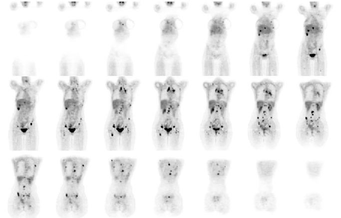
Coronal FDG-PET images.
View main image(pt) in a separate image viewer
View second image(pt).
Anterior and posterior reprojection (volume rendered) FDG-PET views.
View third image(bs).
Whole body bone scintigraphy from an outside hospital.
Full history/Diagnosis is available below
Diagnosis: Metastatic Breast Carcinoma
Full history:
27 year-old woman initially diagnosed with invasive ductal carcinoma by ultrasound guided biopsy. She underwent bilateral mastectomy, chemotherapy, and right-sided radiation.
Radiopharmaceutical:
15.0 mCi F-18 Fluorodeoxyglucose i.v.
Findings:
FDG-PET imaging demonstrates innumerable areas of increased uptake consistent with diffuse metastatic disease.
The whole-body bone scintigraphy study initially performed for right rib pain demonstates focal mildly increased activity in the superior right parietal skull, right lower sternum, left anterior 6th rib, right aspect of L4, superior right acetabulumamd medial proximal right femur.
Discussion:
FDG-PET scanning is a valuable tool to image aggresive neoplasms such as breast cancer. Evaluation of extent of disease is crucial to therapy and overall prognosis. Bone scintigraphy likely significantly underestimated the extent of skeletal metastases.
ACR Codes and Keywords:
References and General Discussion of PET Tumor Imaging Studies (Anatomic field:Breast, Category:Neoplasm, Neoplastic-like condition)
Search for similar cases.
Edit this case
Add comments about this case
Return to the Teaching File home page.
Case number: pt085
Copyright by Wash U MO

