Case Author(s): Jayson R. St. Jacques, M.D. and Tom R. Miller, M.D., Ph.D. , 10/18/02 . Rating: #D3, #Q3
Diagnosis: Gastric Lymphoma
Brief history:
49 year old woman with abnormal upper endoscopy.
Images:

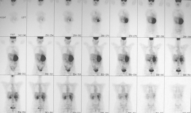
Coronal FDG-PET images.
View main image(pt) in a separate image viewer
View second image(pt).
Coronal FDG-PET images one year later.
View third image(ct).
Axial abdominal CT images before and after therapy.
Full history/Diagnosis is available below
Diagnosis: Gastric Lymphoma
Full history:
49 year old woman with initial presentation of dyspepsia, followed by upper endoscopy and biospy of ulcerated mass. Biospy demonstated B-cell lymphoma. Initial PET scan was for staging, second PET scan was for evaluation of response to chemotherapy.
Radiopharmaceutical:
3.0 mCi F-18 Fluorodeoxyglucose i.v.
Findings:
The initial FDG-PET scan (9-26-01) demonstrates lymphoma confined to the stomach without focal or distant metastatic disease. The differential diagnosis without a known primary malignancy includes adenocarcinoma, lymphoma, active inflammatory/infectious gastritis, and possibly sarcoid or metastatic disease.
The repeat FDG-PET scan (8-20-02) demonstrates complete resolution of abnormal uptake after therapy. Benign bilateral muscle uptake in the lower neck and upper chest was noted.
Discussion:
FDG-PET scanning is excellent for evaluation of lymphoma.
FDG-PET is a useful tool in the treatment of lymphoma for initial staging and post-therapy evaluation. FDG-PET can also be utilized during therapy for early assessment of the response to therapy.
ACR Codes and Keywords:
References and General Discussion of PET Tumor Imaging Studies (Anatomic field:Gasterointestinal System, Category:Neoplasm, Neoplastic-like condition)
Search for similar cases.
Edit this case
Add comments about this case
Return to the Teaching File home page.
Case number: pt084
Copyright by Wash U MO

