Case Author(s): Christian T. Schmitt, M.D. and Farrokh Dehdashti, M.D. , 9/1/02 . Rating: #D2, #Q3
Diagnosis: Poorly differentiated thyroid carcinoma
Brief history:
64 year-old man with a cough and an enlarging right neck mass
Images:
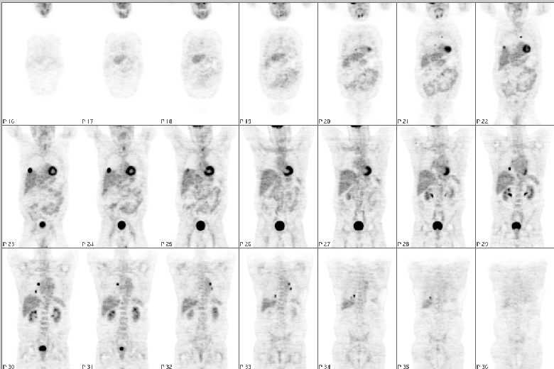
Coronal images are shown
View main image(pt) in a separate image viewer
View second image(tr).
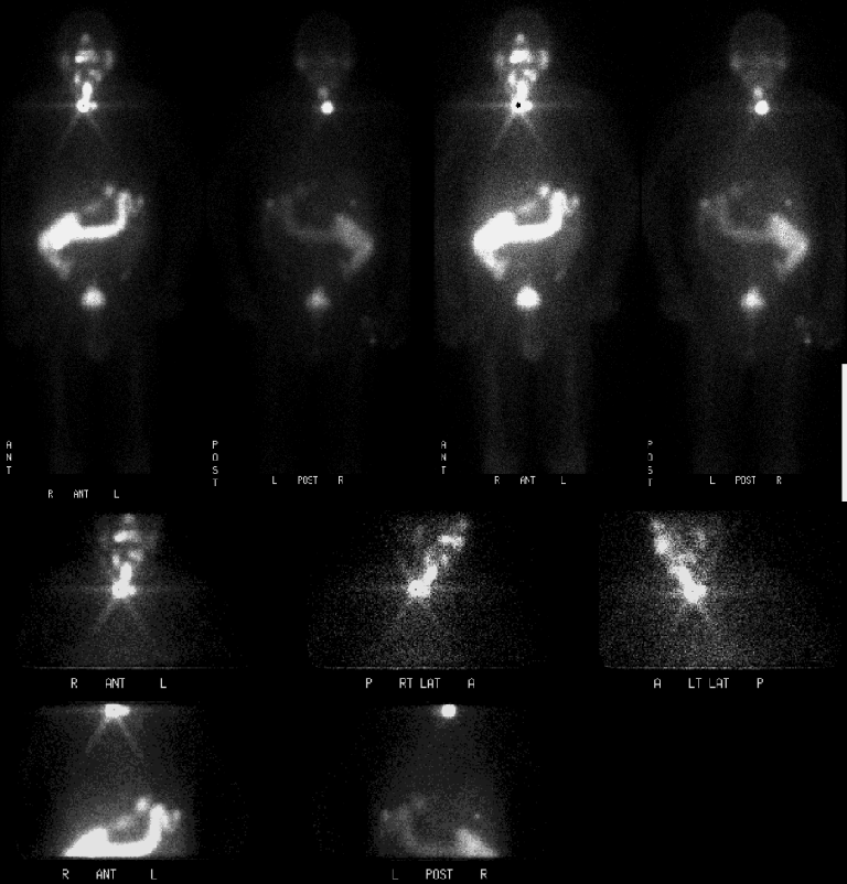
Anterior and posterior images from a second study
View third image(ct).
Selected axial images from initial study obtained
View fourth image(pt).
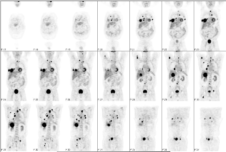
Follow up study, coronal images are shown
Full history/Diagnosis is available below
Diagnosis: Poorly differentiated thyroid carcinoma
Full history:
64 year-old man who initially presented in January 2000 with a cough and an enlarging right neck mass. CT revealed a thyroid mass extending into the superior mediastinum. FNA biopsy revealed neuroendocrine epithelium favoring a follicular thyroid lesion. The patient underwent thyroidectomy and at the time of surgery, the mass extended into the anterior mediastinum. There were several satellite nodules and 1/6 neck nodes were positive for malignancy. There was vascular invasion as well. The surgical margins were positive as well. Pathology indicated a poorly differentiated thyroid carcinoma. The patient received 250mCi of I-131 following thyroidectomy. He also received chemotherapy with Adriamycin, and external beam radiation therapy. The patient initially had a FDG PET study for staging and subsequently a post treatment I-131 whole body study. The patient's thyroglobulin level had continued to rise as shown in the table below. The patient returned with symptoms of diplopia attributable to the left eye, right thigh pain and a lump on the superior aspect of the sternum. The second FDG PET study was performed for restaging.
Radiopharmaceutical:
F-18 Fluorodeoxyglucose as well as I-131 Sodium Iodide
Findings:
Initial CT (3/22/00): Right sided mass extending from thyroid into superior mediastinum.
Initial FDG PET (5/25/00): Numerous lung metastases.
Post-treatment I-131 Scan (5/30/00): No I-131 uptake in known lung metastases.
FDG PET (8/26/02): Interval progression with diffuse pulmonary, hepatic, osseous, and soft tissue metastases as well as a left orbit lesion (only partially within field of view).
| Initial PET Scan | Follow up PET Scan |
 |  |
CT Orbits and Chest (8/30/00): (Follow up image listed below) Large left intraorbital mass that is predominantly intraconal. Expansile lytic rib lesions (not shown), multiple bilateral pulmonary nodules and low attenuation liver lesions consistent with metastases.
Discussion:
The initial staging in this patient demonstrates how metastases from poorly differentiated thyroid carcinoma may be identified on FDG PET and not have significant I-131 uptake. Since this patient had pulmonary metastases on FDG PET that did not show I-131 uptake, the patient received chemotherapy and external beam radiation as well as I-131. FDG PET can also be helpful in patients with a negative I-131 whole body scan in a patient even with more well differentiated thyroid cancer and a rising thyroglobulin level. This patient demonstrated a rising thyroglobulin level as well as the disease progressed.
FDG PET is approved by the CMS for follow up of patients with thyroid carcinoma with an elevated thyroglobulin level and a negative or equivocal I-131 whole body scan.
referrences:
Chung et al., J Nucl Med 1999; 40:986-992.
Helal et al., J Nucl Med 2001; 42:1464-1469.
View followup image(ct).
Selected axial and coronal images from follow up CT
ACR Codes and Keywords:
- General ACR code: 23
- Face, Mastoids, and Neck:
2.373 "Primary carcinoma"
References and General Discussion of PET Tumor Imaging Studies (Anatomic field:Face, Mastoids, and Neck, Category:Neoplasm, Neoplastic-like condition)
Search for similar cases.
Edit this case
Add comments about this case
Return to the Teaching File home page.
Case number: pt083
Copyright by Wash U MO
