Case Author(s): Christian T. Schmitt, M.D. and Farrokh Dehdashti, M.D. , 8/31/02 . Rating: #D3, #Q4
Diagnosis: Urachal Adenocarcinoma
Brief history:
34 year-old man with a one year history of intermittent gross hematuria.
Images:

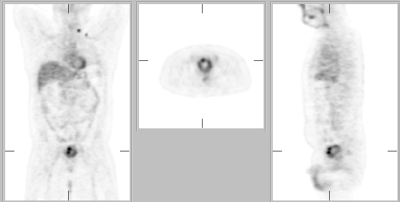
Coronal images are shown
View main image(pt) in a separate image viewer
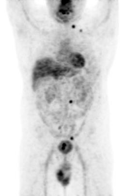
View second image(pt).
Selected coronal, axial and sagittal images are shown
View third image(pt).
Projection images are shown (large file - 1.5MB)
View fourth image(ct).
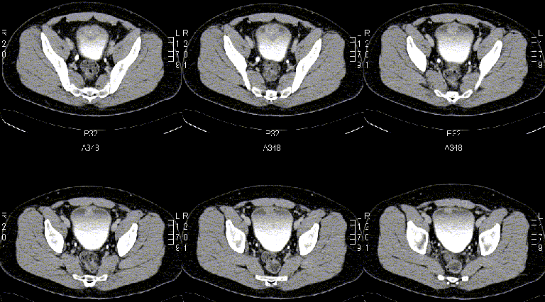
Selected axial CT images are shown
Full history/Diagnosis is available below
Diagnosis: Urachal Adenocarcinoma
Full history:
34 year-old man with a one year history of intermittent gross hematuria initially presented to the ER for urinary retention probably secondary to taking a lot of cold medicine. An intravenous urogram was performed (not shown) demonstrating moderate trabeculation of the bladder. Subsequent cystoscopy demonstrated a large (3-4cm) bladder tumor on the posterior wall and dome. The patient underwent a transurethral resection and presented for FDG-PET imaging to assist staging.
Radiopharmaceutical:
F-18 Fluorodeoxyglucose
Findings:
FDG PET: Increased FDG uptake in lymph nodes in the left supraclavicular, left para-aortic, right common iliac and probable left external iliac regions compatible with metastases. Two foci of increased FDG uptake along the anterior wall of the bladder.
CT Abdomen and Pelvis: Soft tissue lesion in the anterosuperior bladder wall. Enlarged lymph node in the left para-aortic region.
Although, FDG-PET is limited in evaluation of bladder tumors due to excretion of FDG in the urine, it is very useful for determination of metastatsis to lymph nodes or distant organs.
Reference:
Hofer et al., Eur Urol 2001: 40:481-487.
Discussion:
Urothelial neoplasms, such as transitional cell carcinomas are much more common than adenocarcinomas of the bladder. Adenocarcinomas may be primary, related to a persistent urachal remnant, or metastatic to the bladder. Adenocarcinoma is the most common type of urachal carcinoma. The prognosis is poorer than that of transitional cell carcinoma, and it is related to the pathological stage and grade at presentation, and lymph node involvement. Urachal adenocarcinomas can invade locally or metastasize to regional lymph nodes or spread further to the lungs, bones or liver. Surgical resection with wide margins is the preferred treatment. Chemotherapy and radiation have not been shown to be very effective.
Followup:
Initial resection of the bladder tumor revealed a poorly differentiated adenocarcinoma invasive into the muscularis propria. The immunohistochemical findings were compatible with a urachal adenocarcinoma. CT-guided biopsy of the left para-aortic lymph node revealed poorly differentiated adenocarcinoma.
View followup image(ct).
Subsequent CT-guided biopsy of the retroperitoneal left para-aortic lymph node.
ACR Codes and Keywords:
- General ACR code: 83
- Genitourinary System:
8.32 "MALIGNANT NEOPLASM-PRIMARY"
References and General Discussion of PET Tumor Imaging Studies (Anatomic field:Genitourinary System, Category:Neoplasm, Neoplastic-like condition)
Search for similar cases.
Edit this case
Add comments about this case
Return to the Teaching File home page.
Case number: pt082
Copyright by Wash U MO