Case Author(s): Christian T. Schmitt, M.D. and Keith Fischer, MD , 8/28/02 . Rating: #D2, #Q3
Diagnosis: Non-Hodgkins Lymphoma
Brief history:
66 year old HIV-positive man with anemia and fever of unknown origin
Images:

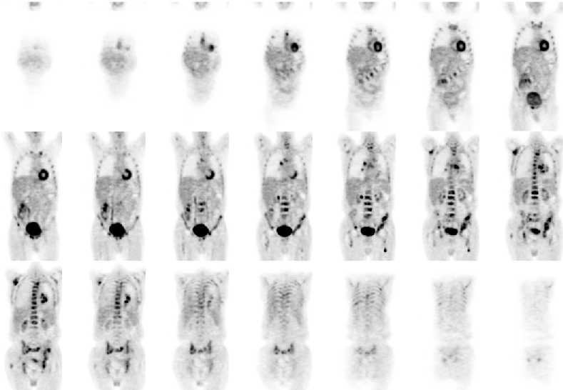
Coronal images are shown
View main image(pt) in a separate image viewer
View second image(pt).
Projection images are shown (large file - 1.5MB)
View third image(ct).
Selected images from CT
Full history/Diagnosis is available below
Diagnosis: Non-Hodgkins Lymphoma
Full history:
66 year-old man with a known history of HIV complicated with Non-Hodgkins lymphoma presented with fatigue, anemia and a fever of unknown origin. He had previously received 6 cycles of CHOP chemotherapy and presented for evaluation for recurrence and restaging.
Radiopharmaceutical:
F-18 Flurodeoxyglucose (FDG)
Findings:
PET (7-8-02): Diffuse increased FDG activity in the skeleton, including: cervical, thoracic, and lumbar spine, multiple ribs, pelvis, sacrum, proximal femurs, right scapula and sternum. Increased FDG uptake is also noted in multiple bilateral hilar, right paratracheal and bilateral supraclavicular lymph nodes and in the left lower lobe. Compatible with diffuse metastatic lymphoma.
CT of Chest/Abdomen/Pelvis (6-17-02): Several small mediastinal lymph nodes (largest 1.2x0.6cm in aorticopulmonary window) unchanged from prior of 4-15-02. Patchy infiltrate in the left lower lobe. Slight interval increase in retroperitoneal soft tissue from prior. (Low attenuation foci in the spine and pelvis are noted on bone windows in retrospect.)
Discussion:
FDG PET is useful in the initial staging, restaging and for assessment of response to therapy in both Hodgkins and non-Hodgkins lymphoma. It is able to show clinically occult skeletal or bone marrow involvement. CT scans often show residual soft tissue in areas of involvment and cannot always discriminate between treated disease and tumor recurrence. FDG uptake in such areas is usually indicative of recurrence and the absence of FDG uptake nearly excludes residual or recurrent tumor. Therefore, FDG PET can be used to guide the therapy of these patients.
Followup:
The patient had a bone marrow biopsy following the PET study and was subsequently admitted for further evaluation. The initial bone marrow aspirate of the right iliac crest demonstrated hypercellular marrow with multiple granulomas with a focus of necrotic marrow worrisome for, but not diagnostic of involvement by lymphoma. No organisms were found. The bone biopsy of the right iliac bone and the L2 vertebral body on 7-22-02 was negative for lymphoma. The bone marrow biopsy on 7-29-02 from the left posterior iliac crest demonstrated hypercellular marrow with trilineage hematopoiesis and multiple lymphohistiocytic aggregates, but flow cytometry was negative for lymphoma. The patient was started on Epogen for his anemia.
He returned for persistent fevers on 8-26-02 and repeat CT demonstrated interval development of marked mediastinal and left hilar adenopathy and interval collapse of the left lower lobe. The patient is thought to have recurrent lymhoma at this time.
View followup image(ct).
Follow up CT images (8-28-02) demonstrate marked progression of mediastinal lymphadenopathy and persistent low attenuation lesions in the spine and pelvis.
ACR Codes and Keywords:
- General ACR code: 93
- Vascular and Lymphatic Systems:
9.34 "LEUKEMIA, LYMPHOMA, MYELOMA"
References and General Discussion of PET Tumor Imaging Studies (Anatomic field:Vascular and Lymphatic Systems, Category:Neoplasm, Neoplastic-like condition)
Search for similar cases.
Edit this case
Add comments about this case
Return to the Teaching File home page.
Case number: pt079
Copyright by Wash U MO

