Case Author(s): Yonglin Pu, M.D.,Ph.D. and Tom R. Miller, M.D., Ph.D. , . Rating: #D3, #Q4
Diagnosis: Pulmonary Non-Hodgkin's Lymphoma.
Brief history:
71 year-old female with newly diagnosed lymphoma.
Images:

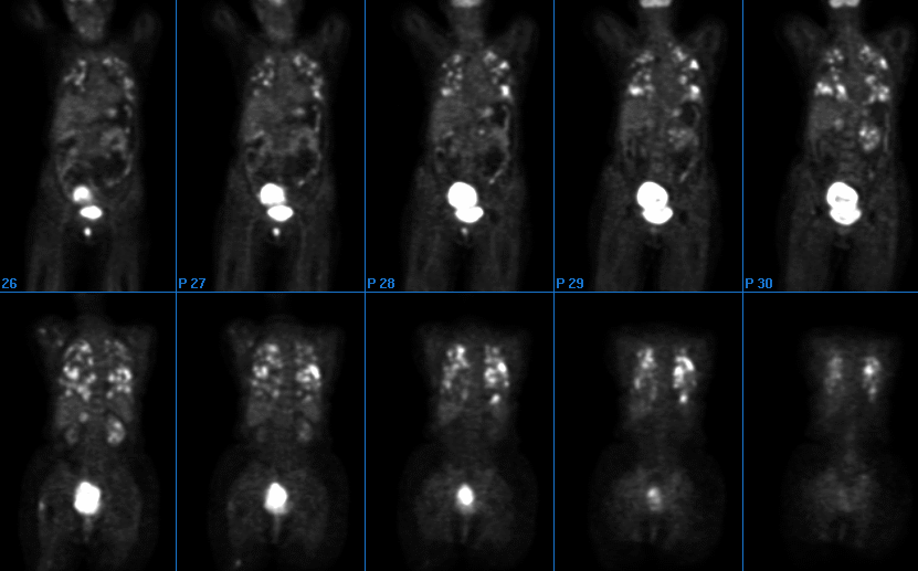
Tumor FDG-PET Imaging
View main image(pt) in a separate image viewer
View second image(ct).
Chest CT one month earlier
Full history/Diagnosis is available below
Diagnosis: Pulmonary Non-Hodgkin's Lymphoma.
Full history:
Newly diagnosed large cell lymphoma involving the lungs, right adrenal gland, left kidney, and a large pelvic mass. The patient had one dose of chemotherapy one day before the PET scan and now presents for staging.
Radiopharmaceutical:
13.5 mCi F-18 Fluorodeoxyglucose
Findings:
Innumerable areas of intense uptake are noted in pulmonary nodules compatible with pulmonary lymphoma. This finding correlates with multiple pulmonary nodules demonstrated on recent CT images. No definite mediastinal uptake is noted, although pulmonary activity adjacent to the mediastinum limits evaluation of the mediastinum.
A small focus of mild uptake is noted in the right parotid space. A small focus of moderate uptake is noted in the left orbital or infraorbital region. These may represent lymph nodes with possible metastses. A small focus of uptake is noted in the soft tissue of right shoulder laterally and posteriorly. A small linear focus of uptake is noted in the right chest wall or rib superiorly.
Intense uptake is noted in the region of the known right adrenal mass, compatible with metastatic involvement as suggested by the CT. Multiple foci of increased uptake are noted within the left kidney corresponding to the low-attenuation lesions on the CT scan, compatible with lymphomatous involvement. The right kidney is small. Intense uptake is noted in the large lobulated pelvic mass with a central region of relatively decreased FDG uptake. Small foci of uptake are noted superior to the right greater trochanter of the femur, in the right femoral neck, and posteriorly in the right mid thigh. These may represent lymphomatous involvement.
Discussion:
FDG-PET in lymphoma is used to stage, re-stage and monitor therapeutic response. It is also used in assessing prognosis.
Primary pulmonary non-Hodgkin’s lymphoma is a rare condition, comprising of less than 5% of all primary extra-nodal lymphoma. Diffuse B-cell or indolent follicular lymphoma are the most common types of lymphoma that involve the lungs. The low-grade lymphoma of mucosa-associated lymphiod cells is the most common indolent subtype. This case demonstrated diffuse involvement of the lungs by large cell lymphoma.
References:
1.Murray and Nadel: Textbood of Respiratory Medicine, 3rd ed. W.B. Sauders Company. Page 1456.
2. Czernin J and Phelps M. Positron emission tomography scanning: current and future applications. Annu Rev Med 2002;53:89-112.
Followup:
The diagnosis of large cell lymphoma was confirmed by biopsy and the patient was started chemotherapy.
Major teaching point(s):
Pulmonary lymphoma should be included in the differential diagnosis of multiple pulmonary nodules.
Differential Diagnosis List
Pulmonary metastases from thyroid carcinoma, colorectal carcinoma and melanoma.
ACR Codes and Keywords:
- General ACR code: 63
- Lung, Mediastinum, and Pleura:
6.34 "LEUKEMIA, LYMPHOMA, MYELOMA"
References and General Discussion of PET Tumor Imaging Studies (Anatomic field:Lung, Mediastinum, and Pleura, Category:Neoplasm, Neoplastic-like condition)
Search for similar cases.
Edit this case
Add comments about this case
Return to the Teaching File home page.
Case number: pt075
Copyright by Wash U MO

