Case Author(s): Yonglin Pu, M.D., Ph.D and Barry A. Siegel, M.D. , 06/02/2002 . Rating: #D3, #Q4
Diagnosis: Peritoneal and Hepatic Metastases
Brief history:
72-year-old man with a history of carcinoma at the junction of the decending and sigmoid colon, status post surgery and chemotherapy. He presented with rising CEA. This study was performed for re-staging.
Images:

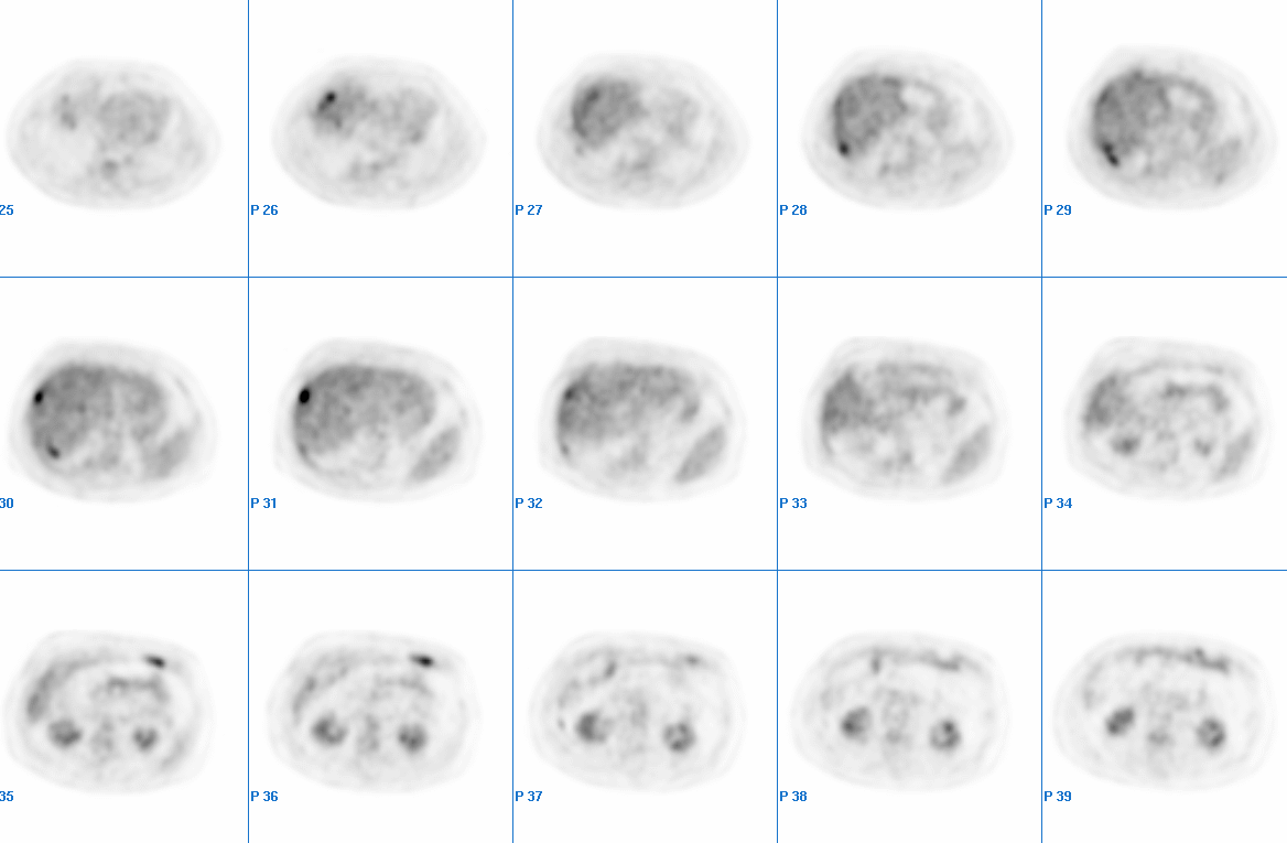
Tumor PET Imaging, Axial image through abdomen
View main image(pt) in a separate image viewer
View second image(ct).
Abdominal CT
Full history/Diagnosis is available below
Diagnosis: Peritoneal and Hepatic Metastases
Full history:
72-year-old man with a history of carcinoma at the junction of the decending and sigmoid colon, status post surgery 2 years ago and chemotherapy completed 14 months ago. He presented with a rising CEA level (most recently 25.2). This study was performed for re-staging.
Radiopharmaceutical:
15.0 mCi F-18 Fluorodeoxyglucose i.v.
Findings:
There are several foci (at least 4) of increased FDG uptake on the surface of the right lobe of the liver. The appearance suggests peritoneal deposits on the hepatic surface rather than hepatic metastases. An additional focus of increased uptake along the lateral aspect of the right lobe appears to be an intrahepatic metastasis. There is another area of focally increased activity posterior to the right lobe of the liver that correlates with a soft tissue density along the posterior peritoneal surface adjacent to the posterior margin of the right lobe of the liver on recent CT images. There are no other findings on the CT to correlate with the other areas of focally increased uptake around the liver. There is another focus of increased activity in the left upper quadrant of the abdomen, which correlates with the soft tissue density in the same location on the comparison CT images in the adipose tissue of the peritoneal cavity.
There also is a photopenic area in the left lobe of the liver, which correlates with the cysts demonstrated on the comparison CT images. There is mildly to moderately increased activity in the right precarinal region as well as the postcarinal region in the mediastinum. This may represent granulomatous inflammation, although metastatic lymphadenopathy cannot be completely excluded.
Discussion:
Peritoneal metastases are difficult to diagnose. CT, ultrasonography and MRI are of limited value in diagnosis. Diagnostic laparoscopy and laparoscopic ultrasonography have been shown to be better than these other techniques. However, they are invasive. This case demonstrates the value of FDG-PET relative to CT for the detection of peritoneal metastases and for evaluating patients with unexplained CEA elevation.
References
Flanagan FL, Dehdashti F, Ogunbiyi OA, Kodner IJ, Siegel BA. Utility of FDG-PET for investigating unexplained plasma CEA elevation in patients with colorectal cancer. Ann Surg 1998; 227:319-323.
McDermott R, Dehdashti F, Siegel BA. Positron emission tomography in colorectal cancer. Semin Colon Rectal Surg 2002; 13:127-138.
Rahusen FD, et al. Selection of patients for resection of colorectal metastases to the liver using diagnostic laparoscopy and laparoscopic ultrasonography. Ann Surg 1999; 230:31-37
Major teaching point(s):
FDG-PET is useful in the diagnosis of peritoneal metastases from colon carcinoma.
Differential Diagnosis List
Granulomatous peritonitis, such as tuberculosis, could cause similar findings.
ACR Codes and Keywords:
- General ACR code: 73
- Gastrointestinal System:
7.3 "NEOPLASM, NEOPLASTIC-LIKE CONDITION"
References and General Discussion of PET Tumor Imaging Studies (Anatomic field:Gasterointestinal System, Category:Neoplasm, Neoplastic-like condition)
Search for similar cases.
Edit this case
Add comments about this case
Return to the Teaching File home page.
Case number: pt074
Copyright by Wash U MO

