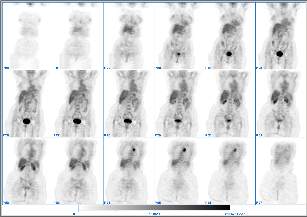
Coronal images of the whole-body FDG-PET.
View main image(pt) in a separate image viewer
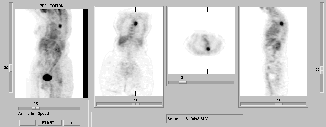
View second image(pt). Selected images of the whole-body FDG-PET.
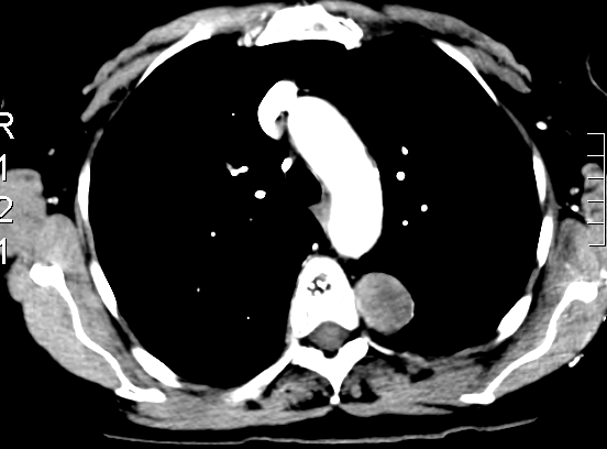
View third image(ct). ct of the chest.
Full history/Diagnosis is available below
CT of the chest: 3.0 x 2.5 cm left paraspinal smoothly marginated mass without osseous changes or neural foraminal widening. The differential diagnosis includes fibrous pleural tumor, lung cancer and, less likely, neurogenic tumor.
This is an example of high fdg uptake in a benign tumor. Other causes of high fdg uptake in the chest of non-malignant etiology: granulamotous inflamation, such as sarcoidosis, histoplasmosis and infectios processes such as bacterial pneumonia.
References and General Discussion of PET Tumor Imaging Studies (Anatomic field:Lung, Mediastinum, and Pleura, Category:Neoplasm, Neoplastic-like condition)
Return to the Teaching File home page.