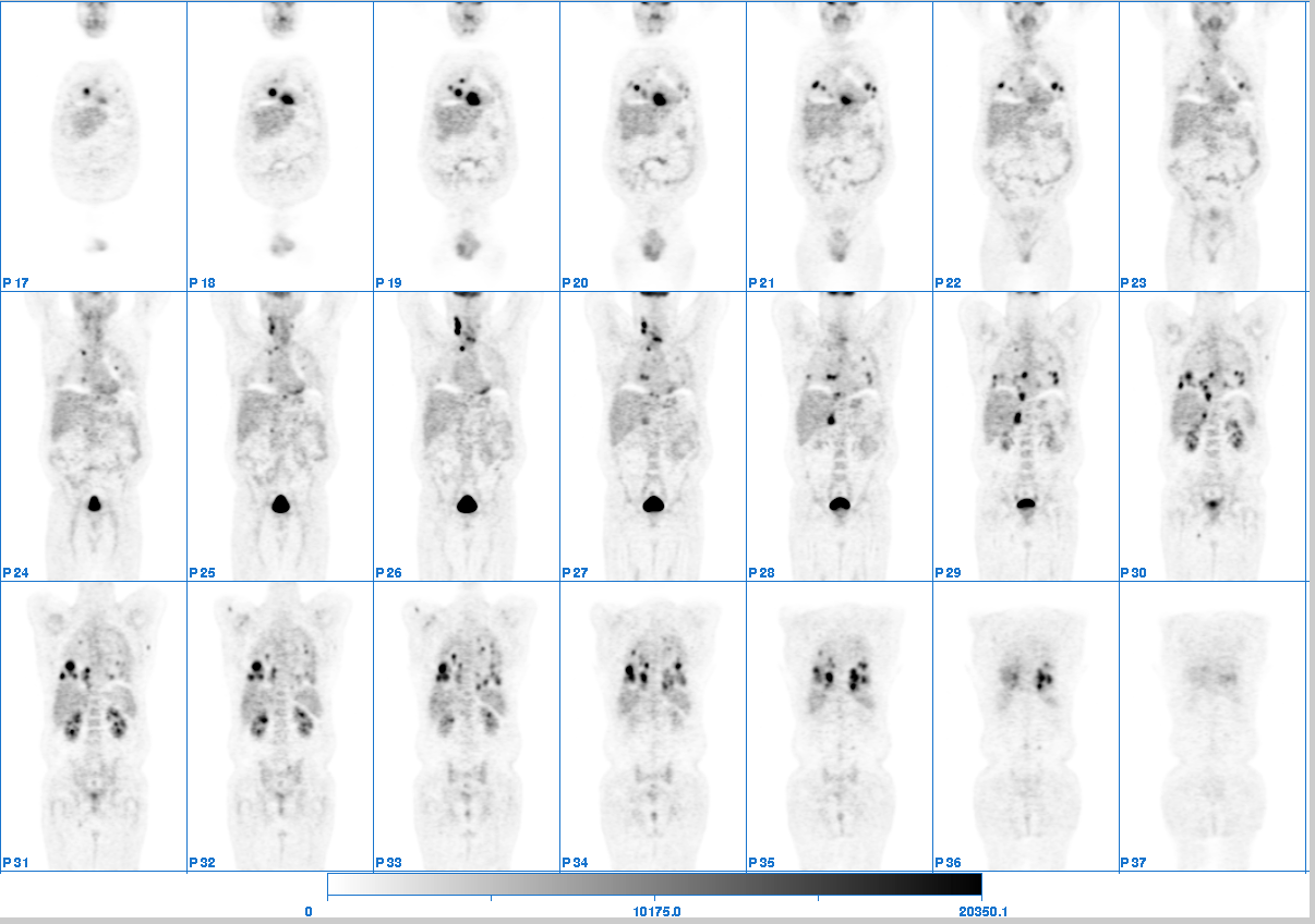
Coronal whole body fdg-pet images.
View main image(pt) in a separate image viewer
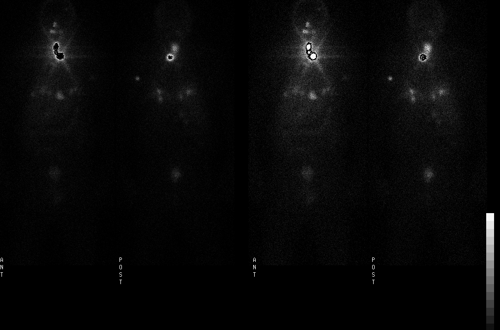
View second image(tr). Whole body images 115 hours after administration of 200 mCi of I-131.
View third image(ct). CT of the chest
View fourth image(ct). CT of the neck
Full history/Diagnosis is available below
I-131 scan: Intense uptake in the region of thyroid gland. There multiple foci of uptake in the lung bases (though some could be in adjacent transverse colon), however, after comparison with CT, not all of the masses evident on CT demonstrate uptake on the I-131 images.
CT-pre-thyroidectomy: Thyroid mass with invasion into the tracheal membrane, with innumerable pulmonary masses.
Selected patients with thyroid cancer can benefit from the use of PET imaging with FDG or with I-124. The PET scan impacts on management by providing (1) more accurate information about staging of patients in terms of extent of tumor for better treatment planning, especially in patients who do not concentration radioactive I-131; (2) the relationship of tumor involvement to vital structures, especially in the neck and central nervous system; and (3) prognostic information (an SUV > 10 and extensive PET + disease connotes a poor prognosis in advanced patients). In the occasional patient, surgically respectable disease has been identified on PET with the result that the patient has been rendered no evident disease with treatment. PET has also been used in the follow-up of patients who have been treated for thyroid cancer, to assess response. PET may also be useful for lesion specific dosimetry, with I-124. The combination of PET and CT in the same gantry facilitates localization of thyroid cancer PET scan abnormalities in relationship to critical organs and structures.
Semin Roentgenol 2002 Apr;37(2):169-74 Positron emission tomography in thyroid cancer management. Larson SM, Robbins R. Department of Radiology, Nuclear Medicine Service, Laurent and Alberta Gershel PET Center, New York, NY, USA.
References and General Discussion of PET Tumor Imaging Studies (Anatomic field:Face, Mastoids, and Neck, Category:Neoplasm, Neoplastic-like condition)
Return to the Teaching File home page.