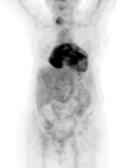

Anterior FDG-PET Projection Image
View main image(pt) in a separate image viewer
View second image(pt). Coronal and Axial FDG-PET Images
View third image(ct). Axial Computed Tomography Images Obtained Six Days Prior to FDG-PET Exam
View fourth image(ct). Axial Computed Tomography Images
Full history/Diagnosis is available below
Axial computed tomographic images (third image) reveal a large pericardial effusion with some thickening and mild enhancement of the pericardium inferiorly.
Axial computed tomographic images (fourth image) through the upper chest demonstrate enlargement and heterogeneous enhancement of the lower pole of the right lobe of the thyroid, corresponding to the FDG-PET abnormality.
One study looking at the use of FDG-PET in suspected soft tissue or osseous infection yielded a sensitivity of 98% and specificity of 75%. Advantages of FDG-PET over other scintigraphic modalities include: rapid diagnosis (2 hours vs. 24-72 hours with Ga-67 or labeled leukocytes), better spatial resolution, the fact that cell-labelling is not required, the ability to evaluate structures that have increased uptake normally on other modalities (eg. liver and spleen), the fact that uptake is less influenced by altered marrow distribution, and the ability to use semiquantitative evaluation. In this case, the inflammatory uptake was noted incidentally.
Incidental thyroid uptake is not infrequently seen on FDG-PET imaging. Often the uptake is diffuse. This can be seen normally or with more significant uptake, in chronic thyroiditis. Occasionally, this can be seen in malignancy, such as lymphoma, so clinical correlation is important. With focal uptake, possibilities include thyroid carcinoma or adenoma. Because experience with focal thyroid uptake is limited, our practice is to recommend thyroid sonography and possible biopsy for further evaluation.
References: Stumpe K et al., Eur J Nucl Med 2000;27:822-832.
Yasuda S et al., Radiology 1998;207:775-778.
The patient underwent ultrasound-guided needle biopsy of the lesion in the right lobe of the thyroid. This demonstrated only hyperplasia.
References and General Discussion of PET Tumor Imaging Studies (Anatomic field:Heart and Great Vessels, Category:Inflammation,Infection)
Return to the Teaching File home page.