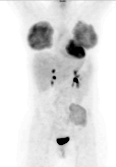
Anterior FDG-PET Projection Image
View main image(pt) in a separate image viewer
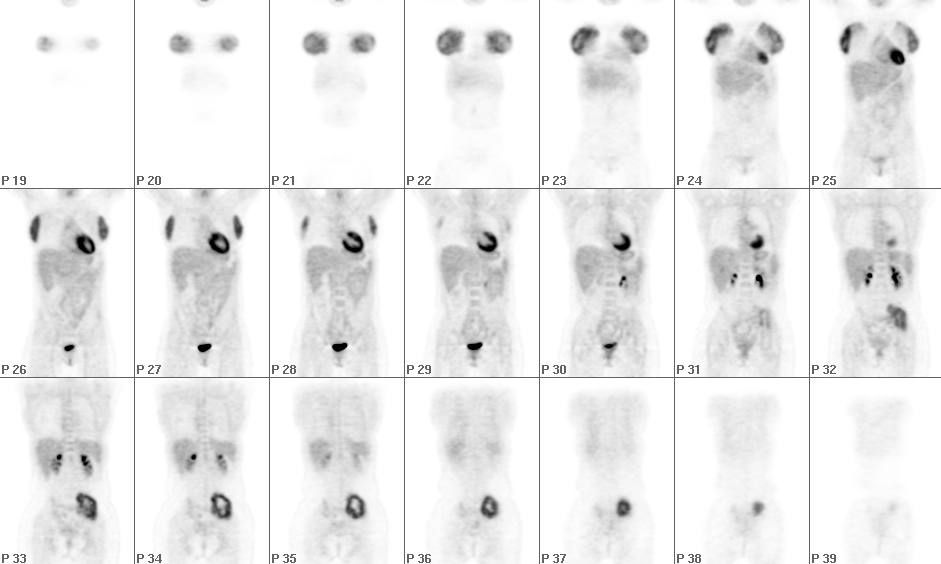
View second image(pt). Coronal FDG-PET Images
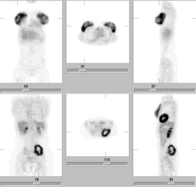
View third image(pt). Coronal, Axial, and Sagittal FDG-PET Images Through the Regions of Interest
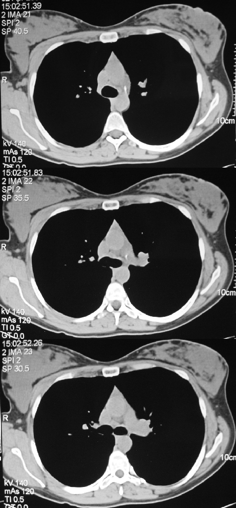
View fourth image(ct). Selected Axial Computed Tomographic Images of the Chest
Full history/Diagnosis is available below
Axial CT images (fourth image) through the thorax demonstrate dense soft tissue in the breasts.
Intense uptake of FDG in lactating breasts has been described. Because the activity is diffuse and bilateral rather than focal, it is unlikely that it would be mistaken for a pathologic process, particularly if the interpreter of the images is aware of the patient's lactation status. Inflammatory breast cancer can cause diffuse FDG uptake, but it would be unusual for it to present bilaterally and symmetrically.
There are limited data regarding dosimetry to an infant due to breast-feeding when the mother underwent FDG-PET imaging. In this patient, minimal activity was found in the breast milk obtained using a breast pump. A 13.4 mL sample of right breast milk obtained at 100 minutes post-injection contained 10.0 nCi/mL/mCi injected decay corrected to the time of sampling. The total volume of this "feeding" represented 0.033% of the injected dose. A 12.4 mL sample of left breast milk obtained 115 minutes post-injection contained 11.2 nCi/mL/mCi injected decay corrected to the time of sampling. The total volume of this "feeding" represented 0.039% of the injected dose.
Thus, the main factor in dosimetry to the infant appears to be the close proximity of the infant to the mother during feeding rather than passage of FDG into breast milk.
References: Schwarzbach MH et al., Clinical value of (18-F) fluorodeoxyglucose positron emission tomography imaging in soft tissue sarcomas. Annals of Surgery. 231(3):380-6, 2000 Mar.
von Schulthess GK, ed. Clinical Positron Emission Tomography: Correlation with Morphological Cross-Sectional Imaging. Philadelphia: Lippincott Williams & Wilkins, 2000.
References and General Discussion of PET Tumor Imaging Studies (Anatomic field:Breast, Category:Metabolic, endocrine, toxic)
Return to the Teaching File home page.