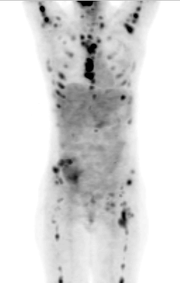
F-18 FDG PET anterior projection image.
View main image(pt) in a separate image viewer
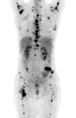
View second image(pt). F-18 FDG PET posterior projection image.
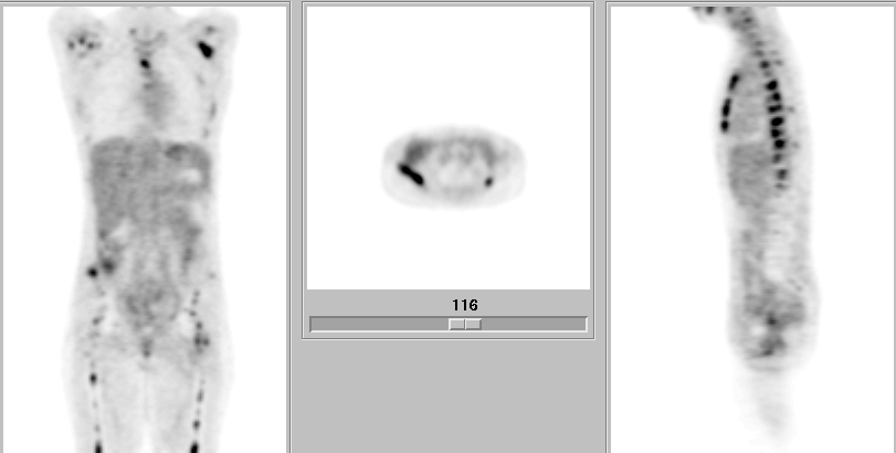
View third image(pt). Multiplanar cross sectional F-18 FDG PET images.
Full history/Diagnosis is available below
A study by Sugawara et al. examined F-18 FDG uptake of histologically proven cervical cancer in 21 patients, revealing a range of SUV’s: 3.68-14.94 (mean 10.31 +/- 3.19).{1} This same study improved visualization of cervical cancers with postvoid images—a concept reinforced by other authors who have sought to minimize urinary activity with bladder irrigation or furosemide post i.v. hydration. Another study by Rose et al. validated the utility of F-18 FDG PET in patients with no CT evidence of extrapelvic disease prior to staging para-aortic lymphadenectomy. Six of eight patients with positive para-aortic node metastases (by histologic analysis) had positive PET scans and there were two false positives, yielding a sensitivity of 75%, specificity of 92%, a PPV of 75%, and a NPV of 92% for predicting disease in CT negative para-aortic lymph nodes. Moreover, one of the two false-negatives had only a microscopic focus of metastatic cancer. Within the pelvis (where nodes could be either CT positive or negative) there were no false-positives or false-negatives resulting in 100% sensitivity, specificity, NPV and PPV. More favorable test characteristics were expected in this location due to the greater prevalence of disease and larger lymph nodes in the pelvis.{2). Grigsby et al. (3), studied 23 women and found that FDG-PET is superior to CT and lymphangiography in demonstrating unsuspected sites of metastasis in pelvic lymph nodes (n=5 patients), in extrapelvic lymph nodes (n=4 patients), and in visceral organs (n=3 patients). PET correctly identified newly diagnosed primary tumor in 10 of 11 patients. In 12 patients with suspected recurrent disease, FDG-PET and CT were positive in 11 and 10 patients, respectively; recurrent disease was confirmed in all 9 patients in whom biopsy was obtained.
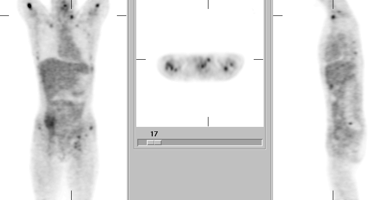
View followup image(pt). Multiplanar F-18 FDG PET images demonstrating supraclavicular and inguinal lymphadenopathy.
Reference: {1} Sugawara Y, Eisbruch A, et al. Evaluation of FDG PET in patients with cervical cancer. Journal of Nuclear Medicine 1999; 40 (7): 1125-31.
{2} Rose P, Adler L, et al. Positron emission tomography for evaluating para-aortic nodal metastasis in locally advanced cervical cancer before surgical staging: a surgicopathologic study. Journal of Clinical Oncology 1999; 17: 41-45.
(3) Grigsby PW, Dehdashti F, Siegel BA. FDG-PET evaluation of carcinoma of cervix. Clinical Positron Imaging 1999; 2:105-109.
References and General Discussion of PET Tumor Imaging Studies (Anatomic field:Genitourinary System, Category:Neoplasm, Neoplastic-like condition)
Return to the Teaching File home page.