Case Author(s): Mark Fister, M.D. and Tom R. Miller, M.D., Ph.D. , 05/17/01 . Rating: #D2, #Q5
Diagnosis: Recurrent CNS Lymphoma
Brief history:
A 62 year-old immunocompetent woman with a history of CNS lymphoma presents with rapid mental deterioration.
Images:
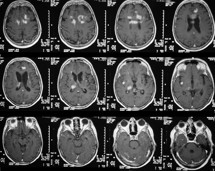
Transaxial contrast-enhanced MR images.
View main image(mr) in a separate image viewer
View second image(pt).
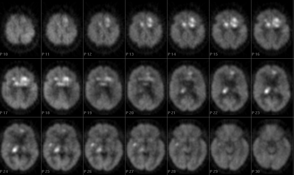
Transaxial F-18 FDG PET images.
View third image(pt).
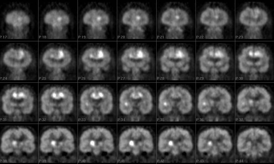
Coronal F-18 FDG PET images.
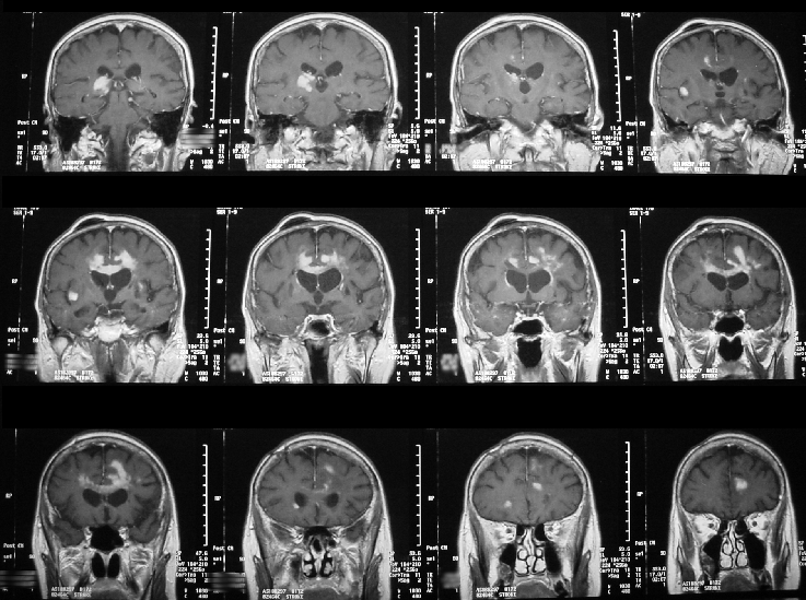
View fourth image(mr).
Coronal contrast-enhanced MR images.
Full history/Diagnosis is available below
Diagnosis: Recurrent CNS Lymphoma
Full history:
A 62 year-old immunocompetent woman with a history of CNS lymphoma presents with rapid mental deterioration. She had been treated eight years ago with radiotherapy and intrathecal chemotherapy. A recent MRI demonstrated new, intensely enhancing multifocal peri-ventricular masses predominating in the frontal lobes. These were felt to represent either radiation necrosis or lymphoma, and PET was recommended for further characterization.
Radiopharmaceutical:
F-18 FDG, i.v.
Findings:
F-18 FDG PET reveals intensely increased metabolism within the enhancing peri-ventricular masses identified by MRI.
Discussion:
Primary CNS lymphoma, though it can occur in immunocompetent patients (particularly the T-cell type), is generally seen in immunocompromised patients, such as patients with AIDS or after organ transplantation. Men are more often afflicted than women, typically in the sixth or seventh decades, and immunocompromised patients are generally affected ten years earlier.
Most CNS lymphomas are supratentorial, predominately involving the deep gray matter or periventricular white matter, and approximately 50% are multifocal. This is in contrast to secondary CNS lymphomas which usually manifest as leptomeningeal involvement. Primary lymphomas can spread over the corpus callosum and can occasionally “coat” the ventricles. They typically exhibit only minimal peritumoral edema. Contrast enhancement is usually moderate although it can be variable, and ring-enhancement may be seen in immunocompromised patients.
CNS lymphomas exhibit dense cellularity and a high proliferative rate, and, therefore, demonstrate high glucose utilization. F-18 FDG uptake is very intense, often suppressing the residual cortical gray matter. This is in contradistinction to nonneoplastic lesions such as toxoplasmosis and radiation necrosis, highlighting PET’s ability to distinguish between these entities.
Followup:
Administration of depot Ara-C via the patient’s indwelling Omaya reservoir and a short course of Decadron, without further radiotherapy, is planned.
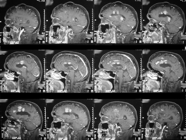
View followup image(mr).
Sagittal contrast-enhanced MRI images.
Major teaching point(s):
Primary CNS lymphomas demonstrates intensely increased uptake on F-18 FDG PET, allowing their differentiation from toxoplasmosis and radiation necrosis
ACR Codes and Keywords:
References and General Discussion of PET Tumor Imaging Studies (Anatomic field:Skull and Contents, Category:Neoplasm, Neoplastic-like condition)
Search for similar cases.
Edit this case
Add comments about this case
Read comments about this case
Return to the Teaching File home page.
Case number: pt058
Copyright by Wash U MO