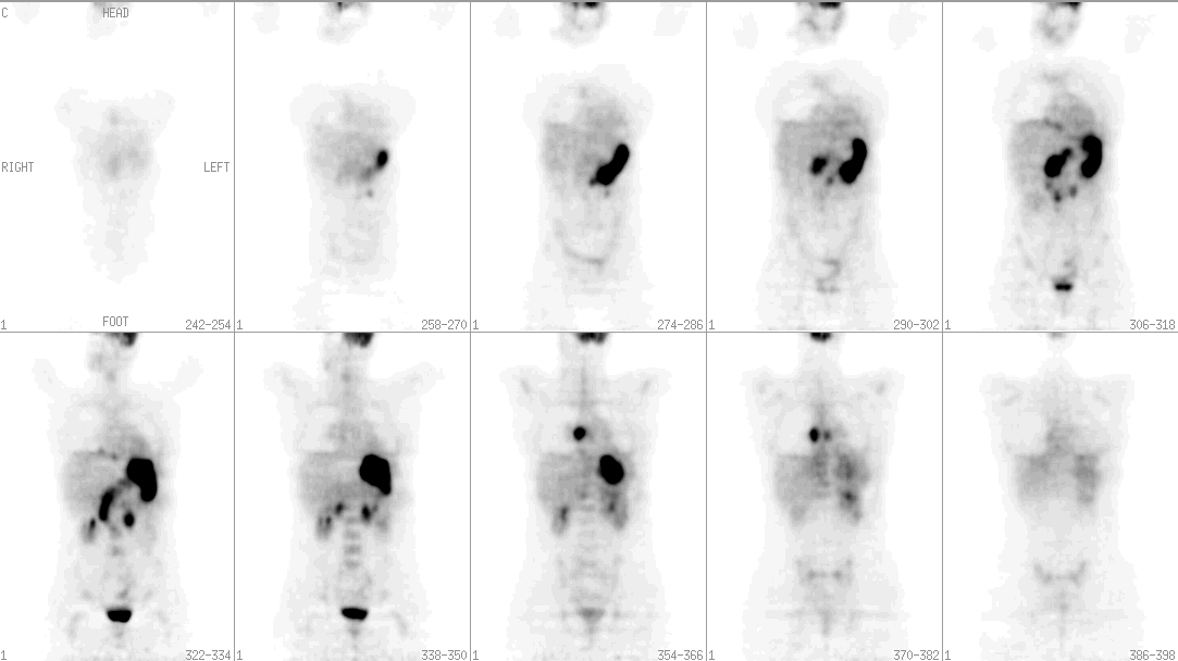
Coronal FDG-PET Images
View main image(pt) in a separate image viewer
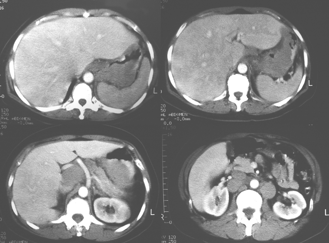
View second image(ct). Selected Axial Computed Tomographic Images of the Abdomen
Full history/Diagnosis is available below
Selected axial CT images (second image) of the abdomen demonstrate marked thickening of the gastric wall with lymphadenopathy predominantly in the upper abdomen.
Advantages of FDG-PET over CT include the fact that anatomic manifestations of disease lag behind metabolic manifestations, and thus FDG-PET has the potential to detect lymphomatous involvement of nodes before they become enlarged. In one study, FDG-PET demonstrated the ability to detect extranodal involvement not seen CT and changed staging in 16% of patients. FDG-PET can often distinguish lymphomatous from non-lymphomatous causes of lymph node enlargement on CT. Following therapy, a residual mass is often seen on CT, which could simply represent scar tissue or could represent lymphoma. These cannot be reliably differentiated with CT. FDG-PET, on the other hand, has shown an accuracy of 90% in distinguishing scar tissue from residual/recurrent lymphoma. Another potential advantage of FDG-PET is the ability to determine early in the course of therapy whether there is response to treatment. Patients could then be spared the debilitating side effects of ineffective chemotherapy and therapy could be altered accordingly.
Advantages of FDG-PET over Ga-67 citrate scintigraphy include better spatial resolution, lower radiation dose, shorter imaging time (approximately 2 hours compared with 48-72 hours) and better evaluation of the abdomen. In addition, FDG uptake is not as dependent on lymphoma cell type as Ga-67 uptake is.
Differential considerations in this patient include gastric adenocarcinoma with lymph node metastases or metastatic disease from another primary malignancy involving the stomach and lymph nodes. Due to the degree of lymph node involvement, lymphoma is the most likely.
Reference: von Schulthess GK, ed. Clinical Positron Emission Tomography: Correlation with Morphological Cross-Sectional Imaging. Philadelphia: Lippincott Williams & Wilkins, 2000.
References and General Discussion of PET Tumor Imaging Studies (Anatomic field:Vascular and Lymphatic Systems, Category:Misc)
Return to the Teaching File home page.