Case Author(s): Yungao Ding, M.D., Ph.D. and Tom R. Miller, M.D., Ph.D. , 03/15/2001 . Rating: #D., #Q.
Diagnosis: Breast cancer with extensive metastases
Brief history:
65 year-old woman with history of back pain and multiple skeletal lesions. Biopsy of a spinal lesion showed metastatic carcinoma of unknown origin.
Images:
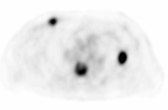
Axial PET image of the chest
View main image(pt) in a separate image viewer
View second image(pt).
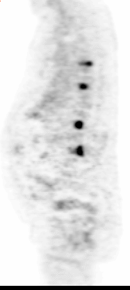
Saggital PET image through the spine
View third image(ct).
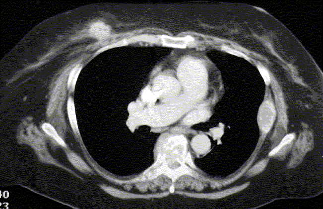
CT of the chest through the breasts
View fourth image(ct).
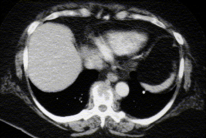
CT of the lower thorax
Full history/Diagnosis is available below
Diagnosis: Breast cancer with extensive metastases
Full history:
65 year-old woman has a history of back pain for more than a year. Prior outside MRI of the spine denostrated multiple spinal lesions. Biopsy of one vertebral lesion showed metastatic carcinoma of unknown origin. The patient has significant bilateral breast skin scarring from a burn at age of 6 which makes mammography difficult to interperate. PET is requested to search for primary cancer.
Radiopharmaceutical:
20 mCi F-18 fluorodeoxyglucose i.v.
Findings:
There is focal, intense uptake of FDG in the right breast as seen in the axial PET image corresponding to the 2 cm right breast mass on the subsequent CT of the chest. The focal intense uptakes of the tracer in T6 thoracic vertebrae and the lateral aspect of the left thoracic wall correspond to destrcuctive vertebral lesions with adjacent soft tissue mass, and the expensile left rib lesion evidenced on the same CT image of the chest through the breasts.
Saggital PET image through the spine demonstrates four spinal lesions with the lower thoracic spinal lesion also detected on CT of the chest.
Additional multiple foci of intense FDG uptakes are also seen on PET (not shown) involving the right internal mammary lymph node chain, bilateral supraaclavicular lymph nodes and lower cervical spine lesions.
Discussion:
The finding of a breast mass with focal intense uptake of FDG in a woman should strongly suggest a breast cancer with or without additional foci of abnormal FDG uptake elsewhere in the body. In this patient with a history of skeletal metastatic carcinoma of unknown origin, the diagnosis of breast carcinoma with systemic metastases is almost certain, although the possibility of the breast lesion being a metastasis cannot be totally ruled out.
Followup:
The right breast lesion was difficult to see on mammography due to significant breast skin scarring bilaterally from burns in childhood. The right breast mass was finally located in the upper inner quadrant after reviewing CT images and was core-biopsied. An invasive ductal carcinoma was diagnosed.
Major teaching point(s):
In a woman with metastatic carcinoma of unknown origin, the possibility of breast cancer should always be strongly considered.
ACR Codes and Keywords:
References and General Discussion of PET Tumor Imaging Studies (Anatomic field:Breast, Category:Neoplasm, Neoplastic-like condition)
Search for similar cases.
Edit this case
Add comments about this case
Return to the Teaching File home page.
Case number: pt054
Copyright by Wash U MO