Case Author(s): Yungao Ding, M.D., Ph.D., Farrokh Dehdashti, M.D. , 03/15/2001 . Rating: #D., #Q.
Diagnosis: Lung cancer with metastases to the abdomen
Brief history:
60 year old woman with newly diagnosed right hilar squamous cell carcinoma
Images:
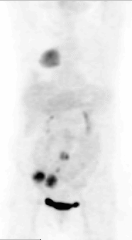
Whole-body reprojection PET imaging
View main image(pt) in a separate image viewer
View second image(pt).
Axial PET image of the lower abdomen
View third image(ct).
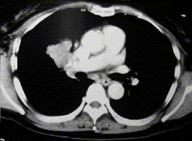
CT of the chest
View fourth image(ct).
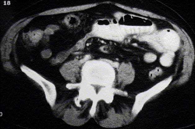
CT of the lower abdomen
Full history/Diagnosis is available below
Diagnosis: Lung cancer with metastases to the abdomen
Full history:
60 year old woman with newly diagnosed right hilar squamous cell carcinoma was evaluated for staging. There is no history of prior malignancy.
Radiopharmaceutical:
14.7 mCi F-18 fluorodeoxyglucose i.v.
Findings:
On PET imaging, there is a large area of intense uptake of FDG in the right hilum corresponding to the large carcinoma demonstrated on CT of the chest.
There are two adjacent regions of intense uptake of FDG in the anterior right lower abdomen. More superiorly, there are two more foci of intense FDG uptake near the midline. Earlier CT of the abdomen and pelvis showed partial small bowel obstruction with dilatation of small bowel to distal ileum but no definite mass in the abdomen or pelvis. There is no retroperitoneal lymphadenopathy. Clinically the patient has vague symptoms related to partial small bowel obstruction.
Discussion:
In this patient with newly diagnosed lung cancer, the findings of multiple areas of intense FDG uptake in the abdomen are suspicious but not typical of metastasis from lung cancer. A second primary malignancy such as colon cancer with mesenteric metastases should also be considered.
Followup:
A colonoscopy was performed and biopsy of the cecum/ileocecal region showed
poorly differentiated carcinoma consistent with adenocarcinoma.
Therefore, a diagnosis of the second primary colon cancer was made.
However, upon surgical resection, the diagosis was changed to squamous
cell carcinoma involving cecum, a loop of small bowel and 3 of 7
resected meseteric lymph nodes. Therefore, this is a case of squamous
cell carcinoma of right hilum with atypical multifocal metastses in the
abdomen. This is an example that PET is superior to conventional
imaging modalities in staging lung cancer. Pieterman et al. have
reported that FDG-PET has a sensitivity of 95% and specificity of 83%
for both mediastinal and distant metastatic disease in non-small-cell
lung cancer (1). In their patient population, PET changed management
in 62 of 102 patients studied.
Reference:
1). Pieterman et al. Preoperative staging of non-small-cell lung cancer with positron emission tomography. New Eng J Med 2000; 343:254-61.
Major teaching point(s):
In patients with known primary cancer, metastasis should always be suspected if malignacy was found in distant locations even if it is not the typical patern of metastasis for that primary acncer.
PET is more sensitive for staging of many cancers such as lung cancer and melanoma.
ACR Codes and Keywords:
References and General Discussion of PET Tumor Imaging Studies (Anatomic field:Lung, Mediastinum, and Pleura, Category:Neoplasm, Neoplastic-like condition)
Search for similar cases.
Edit this case
Add comments about this case
Return to the Teaching File home page.
Case number: pt053
Copyright by Wash U MO