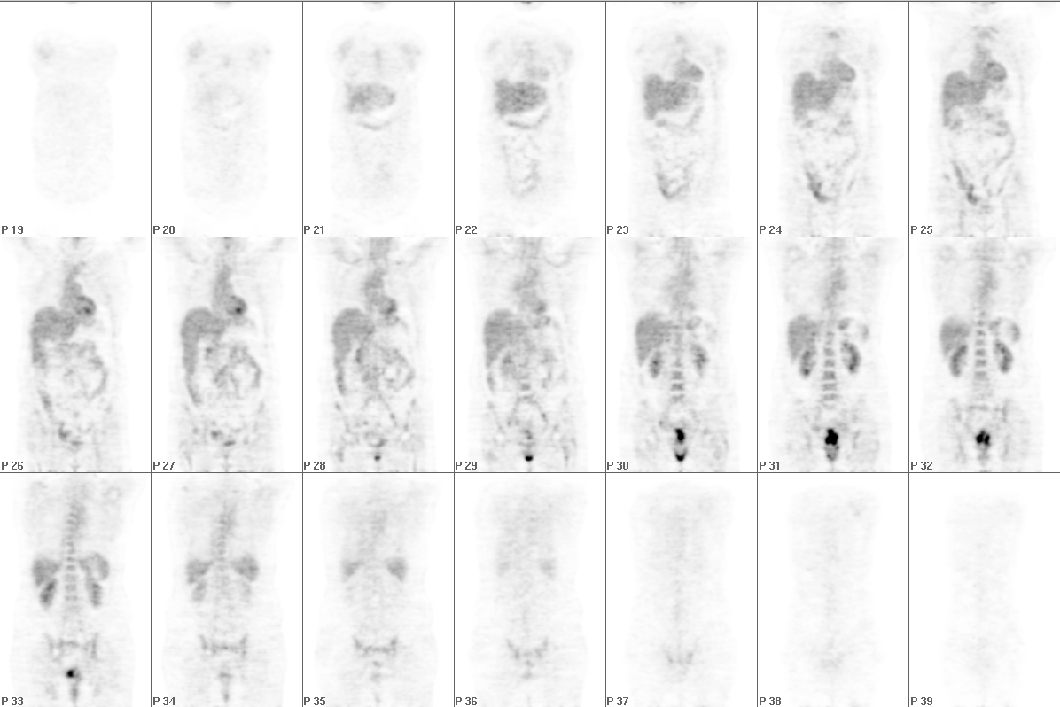
Coronal FDG-PET Images
View main image(pt) in a separate image viewer
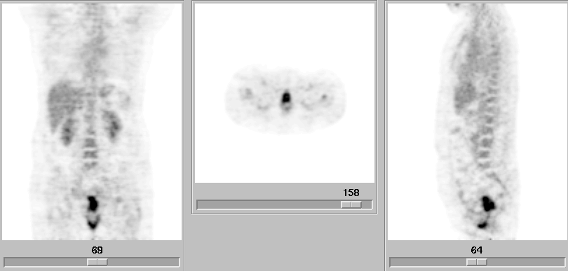
View second image(pt). Coronal, Axial, and Sagittal FDG-PET Images Through the Area of Interest
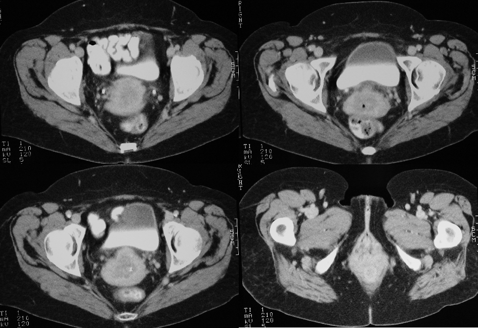
View third image(ct). Selected Axial CT Images
Full history/Diagnosis is available below
Selected CT images (third image) demonstrate a low attenuation mass in the region of the cervix, consistent with cervical cancer. Ill-defined soft tissue fullness is present in the region of the lower vagina.
On physical examination, the patient was noted to have extensive, large condylomata acuminata (not shown) in the lower third of the vagina, corresponding to the region of abnormal uptake on FDG-PET.
FDG-PET has been shown to be useful in the evaluation and staging of cervical cancer, due to the high avidity of cervical cancer for FDG. One study demonstrated uptake in 91% of primary tumors and a sensitivity and specificity for detection of para-aortic nodal metastases of 75% and 92%, respectively. Grigsby et al. have demonstrated that FDG-PET is superior to CT in detection of lymph node metastasis.
FDG uptake can be seen in a number of benign conditions, as well. Increased uptake has been reported in colonic, adrenal and thyroid adenomas, Warthin's tumor, and a number of inflammatory conditions. Given their rapid growth rate, it is not surprising that condylomata acuminata demonstrated increased FDG uptake in this patient.
Other potential causes of this pattern of uptake include extension of tumor, reflux of urine into the vagina (this patient had a foley catheter in place) or inflammation.
References:
Koutsky, L. Epidemiology of Human Papillomavirus Infection. Am J Med. 1997;102(5A):3-8
Rose et al. Positron emission tomography for evaluating para-aortic nodal metastasis in locally advanced cervical cancer before surgical staging: a surgicopathologic study. J Clin Onc. 1999;17(1):41-45
Yasuda S, Ide M, Shohtsu A. Cancer Screening with Whole-Body FDG PET. In: Ruhlmann J, Oehr P, Biersack HJ, eds. PET in Oncology. Berlin:Springer-Verlag, 1999:179-188.
Grigsby et al. PET evaluation of carcinoma of cervix. Clin Pos Img 1999; 2:1050109.
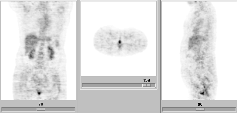
View followup image(pt). Coronal, Axial, and Sagittal FDG-PET Images Through the Area of Interest
References and General Discussion of PET Tumor Imaging Studies (Anatomic field:Genitourinary System, Category:Inflammation,Infection)
Return to the Teaching File home page.