Case Author(s): Stephen Schmitter, M.D. and Tom R. Miller, M.D., Ph.D. , 1/15/01 . Rating: #D3, #Q3
Diagnosis: Crohn's Disease
Brief history:
52 year-old woman with a history of bilateral mastectomies and chemotherapy for breast cancer, who presents for follow-up evaluation.
Images:

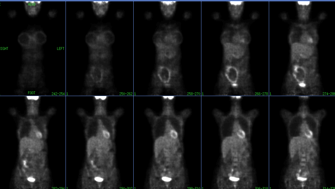
Coronal PET Images
View main image(pt) in a separate image viewer
View second image(pt).
Axial PET Images Through the Lower Abdomen
View third image(ct).
Axial CT Images in the Region of the Cecum
View fourth image(ct).
Axial CT Images Through the Pelvis
Full history/Diagnosis is available below
Diagnosis: Crohn's Disease
Full history:
52 year-old woman with a history of invasive ductal carcinoma and ductal carcinoma in-situ who has undergone bilateral mastectomy and chemotherapy. She had one positive left axillary lymph node at dissection. She now presents for staging evaluation. The patient also has a history of Crohn's disease.
Radiopharmaceutical:
4.3 mCi F-18 Fluorodeoxyglucose i.v.
Findings:
PET images demonstrate areas of decreased uptake in the anterior chest corresponding to breast implants. Mild increased uptake is seen in the left axilla, probably representing post-surgical inflammation. There is no abnormal uptake to suggest recurrent or metastatic disease. There are several foci of markedly increased uptake conforming to bowel in the region of the ileum, cecum and right lower abdomen.
CT images demonstrate several areas of bowel wall thickening in a similar distribution, consistent with the patient's diagnosis of Crohn's disease
Discussion:
Preliminary data suggests that FDG-PET imaging may be useful in evaluating inflammatory processes. In inflammation, activated granulocytes and macrophages undergo a metabolic burst, resulting in increased glucose utilization and thus, increased uptake of FDG.
Increased uptake of FDG has also been observed in patients with inflammatory bowel disease (IBD). Some investigators have suggested a role for FDG-PET as a non-invasive means of monitoring disease activity and response to therapy in patient's with IBD.
Mild to moderate bowel uptake is often noted incidentally in asymptomatic patients. This activity may be related to unrecognized mild inflammation or smooth muscle activity during peristalsis. More intense uptake may indicate clinically significant inflammation or neoplasm and clinical correlation should be pursued.
Reference:
von Schulthess, GK, ed. Clinical Positron Emission Tomography: Correlation with Morphological Cross-Sectional Imaging. Philadelphia: Lippincott Williams & Wilkins, 2000.
ACR Codes and Keywords:
References and General Discussion of PET Tumor Imaging Studies (Anatomic field:Gasterointestinal System, Category:Inflammation,Infection)
Search for similar cases.
Edit this case
Add comments about this case
Return to the Teaching File home page.
Case number: pt046
Copyright by Wash U MO

