Case Author(s): Gabriel De Simon, M.D. and Tom R. Miller, M.D., Ph.D. , 01/05/01. . Rating: #D2, #Q4
Diagnosis: Pulmonary sarcoidosis.
Brief history:
Non-smoker with dry, non-productive cough.
Images:

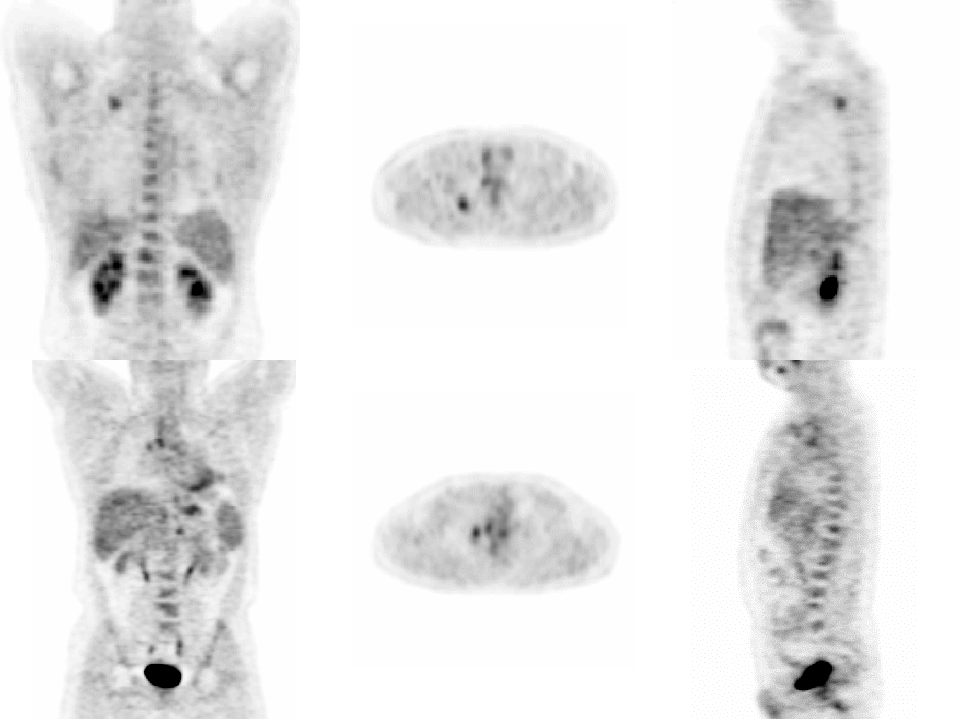
Anterior, posterior, LAO and RAO projection images.
View main image(pt) in a separate image viewer
View second image(pt).
Anterior, transaxial and sagittal images.
View third image(ct).
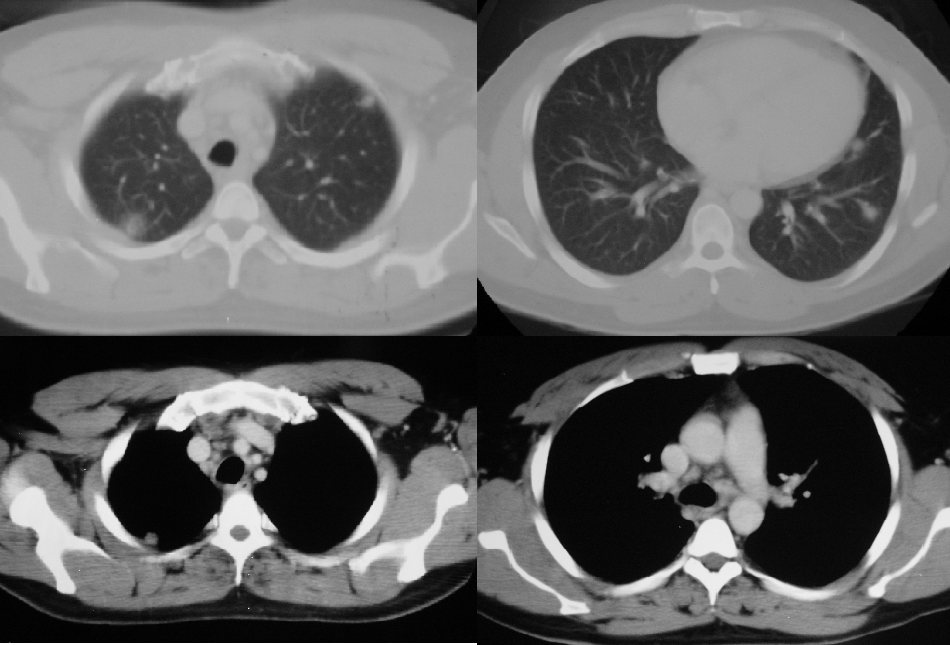
Axial CT images of the thorax.
Full history/Diagnosis is available below
Diagnosis: Pulmonary sarcoidosis.
Full history:
Thirty-four year old construction worker with non-productive cough, in whom a routine chest radiograph demonstrated a questionable right upper lobe mass. The subsequent chest CT showed numerous bilateral pulmonary nodules. The study was requested for evaluation of a primary malignancy.
Radiopharmaceutical:
15.0 mCi F18 Fluorodeoxyglucose.
Findings:
WHOLE BODY FDG PET:
1. Moderately increased uptake(standardized uptake value of 3.0), in the largest pulmonary nodule, located in the apico-posterior segment of the right upper lobe. Moderately increased uptake within a lymph node between the innominate artery and superior vena cava at the same level, and within another lymph node immediately posterior to the sternum in the superior mediastinum. There is also uptake in an anterolateral left apical pulmonary nodule located just caudal to the nodules described above. There is uptake in two additional nodules, postero-superior to the left ventricle, and more peripherally in the left lung at the same level.
2. Increased uptake in right paratracheal, bilateral hilar and subcarinal lymph nodes.
CT SCAN OF THE CHEST:
1. Numerous small right and left hilar, right paratracheal and subcarinal lymph nodes, with short axial diameters of less than one centimeter.
2. Numerous pulmonary nodules, the largest abutting the posterior pleural surface of the right upper lobar apical segment, and others in the apical segment of the left upper lobe, lingula, and latero and postero-basal segments of the left lower lobe.
Discussion:
F-18 Fluorodeoxyglucose has been used in positron emission tomography to determine abnormal glucose metabolism in assisting the evaluation of malignancy in patients with abnormalities found by conventional radiography, or in patients with an existing diagnosis of cancer. F18 FDG is taken up by cells in competition with other sugars, and once inside cells, is phosphorylated by hexokinase, resulting in a polar entity that cannot diffuse out of the cell. Dephosphorylation by glucose-6-phosphatase occurs slowly, and the cellular concentration of F18 FDG is therefore representative of the glycolytic activity of exogenous glucose. Uptake of FDG by malignant cells is facilitated by increased expression of glucose transporter molecules at the cell surface, and tumor cells furthermore have increased levels and activity of hexokinase, and a relative lack of glucose-6-phosphatase, allowing much greater accumulation of FDG compared to normal cells. F18 FDG uptake is however, also accelerated during inflammatory processes such as infections, abcesses and granulomatous diseases, including sarcoidosis and tuberculosis. In sarcoidosis, the cellular infiltrate is composed of macrophages, lymphocytes and epitheliod cells, and can involve almost any organ. The lungs are however, frequently affected, and as in this patient, pulmonary manifestations of the disease dominate. The inflammatory reaction is believed to initially involve pulmonary interstitial tissue with formation of non-caseating granulomas, which are characteristic of the disease. Rapidly dividing cells, such as activated inflammatory cells have a high glycolytic activity in order to satisfy high energy demands, and macrophages are known to have high rates of protein secretion and membrane recycling. Pulmonary granulomatous diseases and infection therefore may predispose to localized FDG thoracic uptake, mimicking metastases and limiting the specificity of whole body scans for patients with suspected cancer. Moreover, since both sarcoidosis and lymphomas affect the lymphoid systems throughout the body, the pattern on FDG PET images is non-specific, and cannot differentiate between the two conditions. The role of FDG PET imaging in the diagnosis of sarcoidosis is therefore limited, and early and subtle abnormalities of the lungs frequently missed by conventional CT of the chest, are more accurately evaluated by high resolution CT. In a patient with proven sarcoidosis however, FDG PET may provide additional useful information. PET gives high quality images, with superior resolution and contrast compared to SPECT images, and it is therefore likely that both the extent of involvement and quantification of disease activity, can be more accurately assessed than with Gallium 67 citrate, which in turn has been more sensitive than serum angiotensin-converting enzyme levels for following the course of the disease.
Followup:
Though non-specific, given the patients young age, lack of history of primary malignancy and normal clinical examination, the findings of multiple bilateral pulmonary nodules with associated mediastinal and symmetrical hilar lymphadenopathy on PET images is more suggestive of a granulomatous disorder, or an infectious process. The patient underwent a transbronchial biopsy, with histology demonstrating non-caseating granulomas characteristic of sarcoidosis. A tuberculin purified protein derivative subcutaneous injection performed subsequently demonstrated 15mm induration, suggestive of concurrent tuberculosis, and the patient is to receive empirical tuberculosis therapy following acid-fast bacillus culture results.
Major teaching point(s):
1. Mechanisms of FDG localization in malignant and inflammatory cells.
2. Non-specificity of FDG PET imaging in the distinction of a pulmonary parenchymal inflammatory process from that of malignancy, and potential use instead in following the course or treatment response of sarcoidosis.
ACR Codes and Keywords:
References and General Discussion of PET Tumor Imaging Studies (Anatomic field:Lung, Mediastinum, and Pleura, Category:Inflammation,Infection)
Search for similar cases.
Edit this case
Add comments about this case
Read comments about this case
Return to the Teaching File home page.
Case number: pt045
Copyright by Wash U MO