Case Author(s): Bart Rydzewski, MD, PhD, Tom R. Miller, MD, PhD , . Rating: #D3, #Q3
Diagnosis: Gastric sarcoma
Brief history:
44 y/o female with early satiety.
Images:

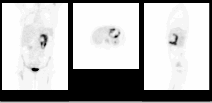
Selected coronal, axial and sagittal FDG PET images
View main image(pt) in a separate image viewer
View second image(pt).
Sequential coronal FDG PET images
View third image(ct).
Axial computed tomography image at the level of the upper abdomen
Full history/Diagnosis is available below
Diagnosis: Gastric sarcoma
Full history:
44 y/o female with a right upper lobe lung mass resected in 1999 and confirmed to represent sarcomatous carcinoma. The patient underwent radiation and chemotherapy treatment to the mediastinum. Currently, she presents with anemia and early satiety. Endoscopic biopsy of the gastric wall demonstrated sarcoma.
Radiopharmaceutical:
15 mCi FDG i.v.
Findings:
There is markedly increased FDG uptake throughout the walls of the stomach. There is no extension of the process into the duodenum or distal esophagus. No other abnormal foci of increased radiopharmaceutical uptake are noted throughout the abdomen and pelvis. There is mildly increased FDG uptake at the apex of the right lung which is likely related to the inflammatory changes due to the right upper lobe mass resection. Computed tomography exam demonstrates a soft tissue mass associated with the wall of the stomach and representing the patient's known sarcoma.
Discussion:
FDG-PET has been demonstrated as a valuable tool in detection and grading of soft tissue and bone sarcomas. Low-grade sarcomas may not be readily detectable, however. In addition, low-grade physiologic uptake in the walls of the alimentary tract needs to be differentiated from malignancy or inflammatory changes. In this case, FDG uptake in the wall of the stomach far exceeded physiologic levels.
Schwarzbach MH. Dimitrakopoulou-Strauss A. Willeke F. Hinz U. Strauss LG. Zhang YM. Mechtersheimer G. Attigah N. Lehnert T. Herfarth C. Clinical value of
ACR Codes and Keywords:
References and General Discussion of PET Tumor Imaging Studies (Anatomic field:Gasterointestinal System, Category:Neoplasm, Neoplastic-like condition)
Search for similar cases.
Edit this case
Add comments about this case
Return to the Teaching File home page.
Case number: pt040
Copyright by Wash U MO

