Case Author(s): Bart Rydzewski, MD, PhD, Tom R. Miller, MD, PhD , 12/05/00 . Rating: #D3, #Q3
Diagnosis: Cervical cancer
Brief history:
57 year old woman with history of cervical cancer
Images:

Selected coronal, axial and saggital images from FDG-PET study.
View main image(pt) in a separate image viewer
View second image(pt).
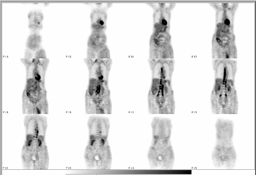
Sequential coronal images from FDG-PET study.
Full history/Diagnosis is available below
Diagnosis: Cervical cancer
Full history:
57 year old woman with history of cervical cancer and retroperitoneal lymph node metastasis, status post radiation therapy 2 years ago. The patient presents with one-month history of left lower extremity swelling. Evaluate for recurrent cervical cancer.
Radiopharmaceutical:
15 mCi FDG i.v.
Findings:
There is extensive bilateral uptake of FDG following the expected course of the iliac vessels in the upper pelvis and extending superiorly adjacent to the lumbar spine and thoracic spine bilaterally to the level of the expected aortic arch. The findings are consistent with extensive metastases along the lymphatic chain. There is a small focal area of increased uptake in the left supraclavicular region and a small area of mildly increased uptake in the left paratracheal region near the level of the clavicle suspected for metastasis. There is no focal area of abnormal uptake of the FDG in the lower pelvis or inguinal regions.
There is a large area of mildly increased uptake in the left thigh along the expected course of the vascular bundle in the soft tissue medially which has an indistinct border. The inferior border of this
area of increased uptake is not imaged and is outside the field of view. The findings are probably due to inflammatory changes such as cellulitis/phlebitis and the possibility of metastasis in the left thigh is unlikely although not totally excluded.
Discussion:
Cervical cancers of both squamous and non-squamous histologies are avid take up FDG, and PET-FDG scanning predicts both the presence and absence of pelvic and para-aortic nodal metastatic disease. One of the limitations of PET scanning of the pelvis with FDG is excreted FDG activity in the urine, although with the advent of PET/CT the problem is somewhat less severe. Typically a Foley catheter is placed and 20 mg of furosemide is administered. Several studies have shown the superiority of FDG-PET over CT in this clinical seting.
Grigsby PW, Siegel BA, Dehdashti F. Lymph node staging by positron emission tomography in patients with carcinoma of the cervix. J Clin Oncol 2001; 19:3745-3749.
Wright JD, Dehdashti F, Herzog TJ, Mutch DG, Huettner PC, Rader JS, Gibb RK, Powell MA, Gao F, Siegel BA, Grigsby PW. Preoperative nodal staging of early stage cervical carcinoma by FDG-PET. Cancer 2005; 104:2484-2491.
Differential Diagnosis List
Lymphoma, acute inflammatory process
ACR Codes and Keywords:
References and General Discussion of PET Tumor Imaging Studies (Anatomic field:Genitourinary System, Category:Neoplasm, Neoplastic-like condition)
Search for similar cases.
Edit this case
Add comments about this case
Return to the Teaching File home page.
Case number: pt039
Copyright by Wash U MO