Case Author(s): Bart Rydzewski MD,Ph.D., Barry A. Siegel M.D. , 12/06/00 . Rating: #D3, #Q4
Diagnosis: Cervical carcinoma, Sarcoidosis
Brief history:
35-year-old woman with newly diagnosed cervical cancer.
Images:
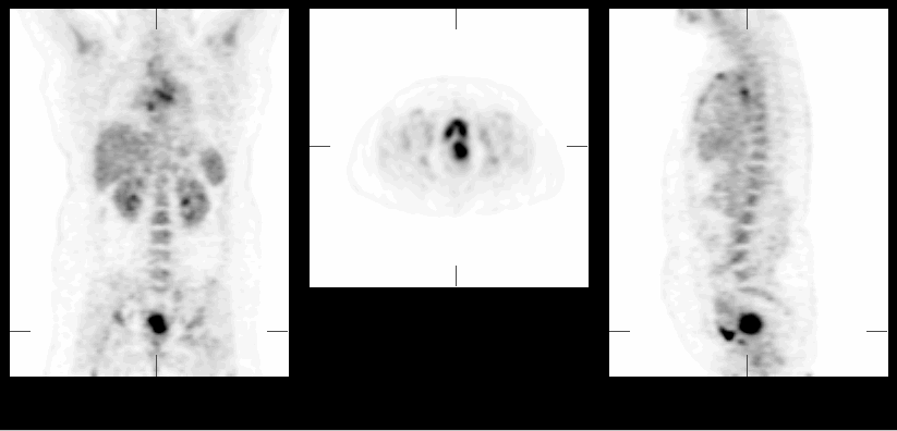
Selected coronal, axial and saggital slices from whole body FDG-PET study
View main image(pt) in a separate image viewer
View second image(pt).
Contiguous coronal slices from whole body FDG-PET study
View third image(ct).
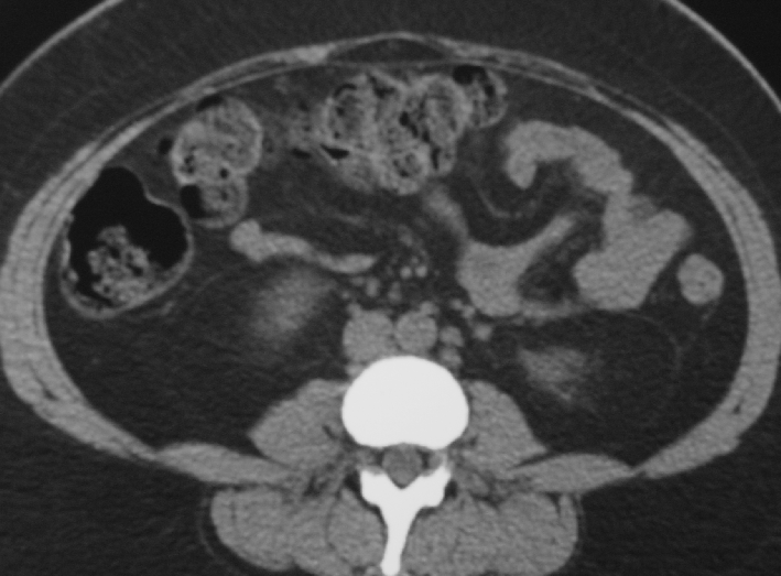
A single axial slice at the level above the iliac crest from a CT examination of the abdomen and pelvis.
Full history/Diagnosis is available below
Diagnosis: Cervical carcinoma, Sarcoidosis
Full history:
35-year-old woman with cervical carcinoma diagnosed two months ago and currently being staged. The patient has no other relevant medical history.
Radiopharmaceutical:
15 mCi FDG i.v.
Findings:
There is markedly increased radiopharmaceutical uptake in the region of the cervix, consistent with the patient's known cervical cancer. Multiple foci of increased radiopharmaceuticaluptake are also noted in bilateral axillary, left supraclavicular, prevascular, retrotracheal, bilateral inguinal and bilateral iliac nodal regions lymph nodes.
Mild lymphadenopathy was confirmed in the para-aortic, iliac and inguinal regions by a CT exam performed 5 days later.
Followup:
The patient underwent exploratory laparotomy with lymph node dissection. Primary cervical cancer was confirmed histopathologically. Non-caseating granulomatous inflammatory changes conistent with sarcoidosis were noted in multiple lymph nodes.
Major teaching point(s):
Sarcoidosis is one of the inflammatory conditions associated with high FDG uptake and potentially resulting in falsely positive interpretation of FDG-PET studies in patients with primary malignances. In fact, it has been shown that FDG uptake level may reflect "disease activity" in sarcoidosis in quantitative terms (per gram lung tissue).
Brudin LH. Valind SO. Rhodes CG. Pantin CF. Sweatman M. Jones T. Hughes JM. Fluorine-18 deoxyglucose uptake in sarcoidosis measured with positron emission tomography. European Journal of Nuclear Medicine. 21(4):297-305, 1994
ACR Codes and Keywords:
References and General Discussion of PET Tumor Imaging Studies (Anatomic field:Vascular and Lymphatic Systems, Category:Inflammation,Infection)
Search for similar cases.
Edit this case
Add comments about this case
Return to the Teaching File home page.
Case number: pt038
Copyright by Wash U MO