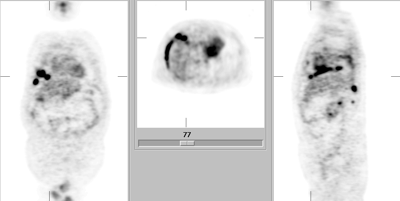

Multiplanar F-18 FDG PET images.
View main image(pt) in a separate image viewer
Full history/Diagnosis is available below
Otherwise occult malignant pleural disease is often suggested indirectly by the presence of a pleural effusion. However, benign effusions related to an underlying tumor, or altogether coincidental effusions also occur in these patients. Traditional evaluation after CT often includes thoracentesis with chemistry (to differentiate transudative vs. exudative effusions) and cytology, and in selected cases, pleural biopsy performed with or without thoracoscopy. Unfortunately, thoracentesis fails to reveal malignancy in approximately one third of malignant effusions, and “closed” pleural biopsy is similarly far from perfect. Thoracoscopic-guided pleural biopsy, though much more sensitive, is a more invasive procedure. As reported by Erasmus, et al., F-18 FDG PET can provide an accurate, noninvasive alternative for characterization of pleural effusions in these patients, with an accuracy similar to that of thoracoscopic biopsy. Using the criterion of pleural uptake greater than mediastinal blood pool, the sensitivity, specificity, PPV, NPV, and accuracy were calculated as 95%, 67%, 95%, 67%, and 92%. Because false-positives occur, Erasmus et al, recommend that malignant pleural disease be confirmed by thoracentesis in questionable cases. In the presence of an exudative effusion, a positive PET, and negative cytology, thoracoscopic-directed biopsy is highly recommended to avoid underdiagnosis and potentially fatal major thoracic surgery.
Reference: Erasmus JJ, McAdams HP, et al. FDG PET of pleural effusions in patients with non-small cell lung cancer. AJR 2000; 175: 245-249.
View followup image(ct). Multiple Axial CT images through the lower thorax.
References and General Discussion of PET Tumor Imaging Studies (Anatomic field:Lung, Mediastinum, and Pleura, Category:Neoplasm, Neoplastic-like condition)
Return to the Teaching File home page.