Case Author(s): Mark Fister, M.D. and Farrokh Dehdashti, M.D. , 9/20/00 . Rating: #D2, #Q5
Diagnosis: Postsurgical abscess.
Brief history:
49 year old man presents for staging after grossly complete excision of a high grade fibrosarcoma from the right groin 1.5 weeks earlier.
Images:

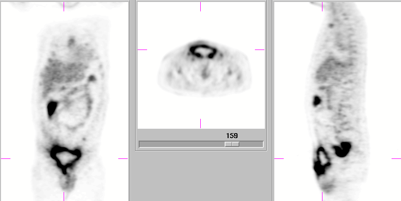
Coronal, Axial, and Sagittal F-18 FDG PET images.
View main image(pt) in a separate image viewer
Full history/Diagnosis is available below
Diagnosis: Postsurgical abscess.
Full history:
49 year old man presents for staging after grossly complete excision of a high grade fibrosarcoma from the right groin 1.5 weeks earlier. Although the surgery was uneventful, the patient had progressively increasing pain at the surgical site following removal of a drain 4 days earlier. He also reported a remote history of rectal cancer, status post abdominoperineal resection 13 years ago, from which he was presumably cured.
Radiopharmaceutical:
F-18 Fluorodeoxyglucose
Findings:
F-18 FDG PET reveals a rim of intensely increased activity surrounding a photopenic region in the anterior abdominal wall at the site of prior surgery. Several small foci of increased uptake adjacent to this region were also noted, as well as increased uptake within two right internal iliac and one left inguinal lymph node.
Discussion:
Although F-18 FDG body PET is conventionally thought as primarily a
means of oncologic imaging, preliminary results at imaging infection
appear promising. This may have greatest significance when considering
the 24-72 hour delayed imaging often required with Ga-67 and In111-WBC
alternatives. Although imaging of infection was inadvertent in this
case, by recognizing the characteristic PET appearance of an abscess
and correlating with the grossly complete excision just 1.5 weeks
earlier, a previously unsuspected abscess was brought to clinical
attention. A similar pattern of peripherally increased FDG uptake
was noted in an abscess reported by Sugawara et al., with the most
intense FDG uptake within the abscess wall, a finding expected with
significant central liquefaction. It should also be noted, that in
oncology patients with significant infection, increased activity within
regional lymph nodes may be either reactive or neoplastic in character.
Repeat PET imaging after complete resolution of infection
is required for reliable characterization of the lymphadenopathy.
Reference: Sugawara Y, Braun DK, et al. Rapid detection of human infections with fluorine-18 fluorodeoxyglucose and positron emission tomography: preliminary results. Eur J Nucl Med 1998; 25: 1238-1243.
Followup:
The patientís surgeon was notified of this unsuspected abscess. After being examined in the surgeonís office, the patient was hospitalized and underwent emergent open drainage of a large abscess.
Major teaching point(s):
Not all increased FDG uptake is due to neoplasm. Infection can demonstrate intensely increased uptake, and the presence of characteristic PET findings in recently postsurgical patients should alert the interpreter to that possibility. Also, the presence of significant infection can confound characterization of regional lymph nodes.
Differential Diagnosis List
1. Postsurgical abscess.
2. Noninfected fluid collection amidst cellulitis.
3. Centrally necrotic tumor.
ACR Codes and Keywords:
References and General Discussion of PET Tumor Imaging Studies (Anatomic field:Skeletal System, Category:Inflammation,Infection)
Search for similar cases.
Edit this case
Add comments about this case
Return to the Teaching File home page.
Case number: pt035
Copyright by Wash U MO

