Case Author(s): Mark Fister, M.D. and Farrokh Dehdashti, M.D. , 09/20/00 . Rating: #D3, #Q4
Diagnosis: Multiple synchronous bronchogenic carcinomas
Brief history:
64 year-old male smoker presents for further evaluation of two right lower lobe (RLL) nodules noted on a chest radiograph obtained as part of a yearly physical.
Images:

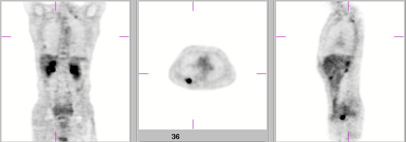
Coronal, Axial, and Sagittal F-18 FDG PET images.
View main image(pt) in a separate image viewer
View second image(ct).
Axial CT through the lower chest.
View third image(ct).
Axial CT of the upper chest.
View fourth image(ct).
Axial CT through the mid chest.
Full history/Diagnosis is available below
Diagnosis: Multiple synchronous bronchogenic carcinomas
Full history:
64 year-old male smoker presents for further evaluation of two right lower lobe (RLL) nodules noted on a chest radiograph obtained as part of a yearly physical. These nodules were confirmed on a subsequent CT, which also revealed a third RLL nodule, numerous granulomata, and biapical scarring. Using CT “lung windows” the RLL nodules measured 1.7 cm diameter, 0.8 x 1.3 cm, and < 3 mm diameter. “Nodular components” of the biapical scarring measured 5 mm diameter, bilaterally.
Radiopharmaceutical:
F-18 Fluorodeoxyglucose, i.v.
Findings:
F-18 FDG PET reveals intensely increased uptake (SUV = 7) within the largest (1.7 cm) RLL nodule. The second largest nodule (0.8 x 1.3 cm) exhibits mildly increased uptake (SUV = 1.4), and was felt to most likely be inflammatory in character, perhaps related to granulomatous infection given the numerous scattered calcified granulomata. The third nodule (< 3m) is not seen on PET. The “nodular” scarring (5 mm) in the right apex demonstrates moderately increased activity (SUV = 2.2) as does that in the left apex (SUV = 1.65). Although the SUV of the apical lesions is within the “benign range”, given their small size, malignancy is suspected, although the specificity of this finding is reduced by the presence of granulomatous disease.
Discussion:
One of the great uses for FDG-PET is the noninvasive characterization of solitary (or multiple, as in this case) pulmonary nodules (SPN). Greater than 50% of SPN’s are benign and could fall victim to invasive attempts at their characterization with attendant complications. The commonly utilized cut-off SUV level of 2.5 for distinguishing malignant from benign lesions must be used cautiously, accounting for susceptibility to partial volume averaging and motion effects. Klemenz and Taaleb claim that for contemporary PET scanners, partial volume and motion effects should be considered in lesions < 2-3 cm in size. They also recommend attenuation correction for small lesions, which could otherwise be missed due to increased background or scatter on uncorrected images. Sources of false-positive results include granulomatous or other infectious processes. False-negatives include the relatively low uptake seen in bronchoalveolar carcinoma, and predominance of scar in cicatricial carcinomas.
Reference: Klemenz B, and Taaleb KM. “Lung cancer” in PET in Clinical Oncology Eds. Coleman RE, and Wieler HJ. Steinkopff Verlag, Darmstadt 2000, pp.193-210.
Followup:
The patient underwent right lower lobectomy which revealed 3 synchronous bronchogenic carcinomas of 3 distinct histologic subtypes. The largest lesion (1.5 cm by gross) was a poorly differentiated adenocarcinoma. The second lesion (1.3 x 1 cm by gross) was a poorly differentiated squamous cell carcinoma. The smallest lesion (0.3 cm by gross) was a bronchoalveolar carcinoma. Followup CT performed 8 months later revealed the “nodular” component of the left apical scarring to be markedly increased in size, measuring 2.9 x 3.8 cm. Subsequent PET revealed markedly increased uptake (SUV = 4.5) in this lesion, as well as moderately increased uptake (SUV = 2.7) in the 5 mm diameter “nodular” scarring at the right apex. CT guided fine needle aspiration of the left apical lesion revealed poorly differentiated non-small cell carcinoma. The character of the right apical lesion remains pathologically unproven.
View followup image(pt).
Coronal and Axial F-18 FDG PET images with correlating CT images.
Major teaching point(s):
Size matters. Partial volume effects should not be ignored in characterizing pulmonary nodules.
Differential Diagnosis List
1. Neoplasm.
2. Granulomatous Infection.
3. Bland scarring.
ACR Codes and Keywords:
References and General Discussion of PET Tumor Imaging Studies (Anatomic field:Lung, Mediastinum, and Pleura, Category:Neoplasm, Neoplastic-like condition)
Search for similar cases.
Edit this case
Add comments about this case
Return to the Teaching File home page.
Case number: pt034
Copyright by Wash U MO

