Case Author(s): Bart Rydzewski M.D., Ph.D. and Farrokh Dehdashti M.D. , 8/23/00 . Rating: #D2, #Q3
Diagnosis: Metastatic melanoma
Brief history:
87 year old male with a history of melanoma of the right forearm.
Images:

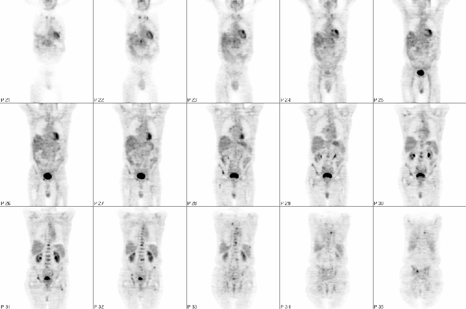
Sequential coronal slices from FDG-PET study
View main image(pt) in a separate image viewer
View second image(pt).
Sequential axial slices from FDG-PET study
View third image(pt).
Selected coronal, axial and saggital slices from FDG-PET study
View fourth image(ct).
Axial slice of CT of the chest exam.
Full history/Diagnosis is available below
Diagnosis: Metastatic melanoma
Full history:
87 year-old male with a history of melanoma of the right forearm which was treated surgically. The patient experienced recurrence with metastatic disease in the right lung and right axilla. Further staging evaluation is requested
Radiopharmaceutical:
15.0 mCi F-18 Fluorodeoxyglucose i.v.
Findings:
There is extensive osseous metastatic disease. Foci of markedly increased uptake are identified in the right and left proximal humeri, the right glenoid region, the left clavicle near the sternoclavicular joint, two posterior right rib lesions (one in the mid hemithorax and the other in the lower right hemithorax), the posterolateral aspect of a left lower rib adjacent to the spleen, multiple lesions in the thoracic, lumbar, and sacral spine, the pelvis bilaterally, and the right and left proximal femurs. Foci of increased uptake are also identified in the right and left aspects of the superior portion of the sternum, which could represent post-surgical change, in this patient with prior median sternotomy; however malignancy involvement in these regions cannot be excluded.
Three nodules are identified in the right lung and one in the left lung. Only 2 of these nodules were seen on CT.
Discussion:
Malignant melanoma has been traditionally staged with combination of
chest radiography, CT, MRI and ultrasoound of the abdomen and axillary,
inguinal and cervical lymph nodes (1-3). These methods however, have
significantly lower sensitivity and specificity for detection of
metastatic foci than FDG-PET. In fact, malignant melanoma shows one
of the highest FDG uptakes of all tumors. Wahl et al. reported (4)that FDG-PET has a sensitivity of 92% and specificity of 100%. Use of FDG-PET in staging of malignant melanoma has been shown to be cost effective (5).
1. Clinical Positron Emission Tomography ed. Schulthess GK, Lippincott Williams & Wilkins 2000
2. Boeni R et al. British Journal of Dermatology 1995;132:556-62
3. Steinert HC et al. Radiology 1995;195:705-9
4. Wahl RL et al. Cancer 1991;67:1544-50
5. Damian DL et al. Melanoma Research 1996;6:325-9
ACR Codes and Keywords:
References and General Discussion of PET Tumor Imaging Studies (Anatomic field:Vascular and Lymphatic Systems, Category:Neoplasm, Neoplastic-like condition)
Search for similar cases.
Edit this case
Add comments about this case
Return to the Teaching File home page.
Case number: pt033
Copyright by Wash U MO

