Case Author(s): Daniel E. Appelbaum, MD and Tom R. Miller, MD, PhD , 6/13/00 . Rating: #D2, #Q5
Diagnosis: Sarcoidosis
Brief history:
Past history of cervical carcinoma with new CT abnormality.
Images:

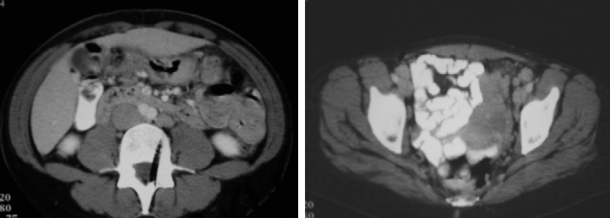
Selected axial images from an abdominal-pelvic CT are shown.
View main image(ct) in a separate image viewer
View second image(pt).
Whole-body FDG-PET PROJECTION images with selected anterior, lateral, and both anterior oblique views.
View third image(pt).
Standard coronal FDG-PET whole body images are shown.
Full history/Diagnosis is available below
Diagnosis: Sarcoidosis
Full history:
36 year-old female with bilateral lower extremity swelling. Ultrasonography was negative and a CT of the abdomen and pelvis was obtained to further evaluate for venous obstruction.
The patient has a past history of localized cervical carcinoma which was treated with a cone resection eleven years ago. (There was no radiation or chemotherapy).
Radiopharmaceutical:
15.0 mCI F-18 Fluorodeoxyglucose (FDG) i.v.
Findings:
The abdominal-pelvic CT demonstrated 1-2cm adenopathy in the retroperitoneal, iliac, and inguinal regions. Given her past history this was suspicious for recurrent cervical carcinoma, although the distribution was somewhat atypical. PET was requested for further evaluation in the hope that the distribution of disease my help distiguish cervical carcinoma from lymphoma or another primary malignancy.
PET images demonstrate massive and extensive adenopathy in the cervical, supraclavicular, axillary, mediastinal, hilar, internal mammary, retroperitoneal, iliac, inguinal, and femoral regions.
Discussion:
While FDG-PET is very sensitive for a variety of malignancies, it can lack specificity. In addition to neoplasm, any active infectious or inflammatory process can demonstrate dramatic FDG avidity. Active histoplasmosis and sarcoidosis can be particularly troublesome when they have an insidious onset of symptoms.
Given the distribution of adenopathy in this case as demonstrated by PET, it was suggested that metastatic cervical carcinoma was extremely unlikely. Lymphoma, sarcoidosis, and disseminated tuberculosis were given as much more likely differential considerations.
Followup:
Biopsy of a cervical lymph node near the base of the neck as localized by PET yielded non-caseating granulomas, compatible with sarcoidosis.
ACR Codes and Keywords:
References and General Discussion of PET Tumor Imaging Studies (Anatomic field:Vascular and Lymphatic Systems, Category:Inflammation,Infection)
Search for similar cases.
Edit this case
Add comments about this case
Return to the Teaching File home page.
Case number: pt031
Copyright by Wash U MO

