Case Author(s): Daniel E. Appelbaum, MD and Tom R. Miller, MD, PhD , 6/13/00 . Rating: #D3, #Q4
Diagnosis: Pelvic inflammatory disease and cervical cancer.
Brief history:
Newly diagnosed malignancy.
Images:
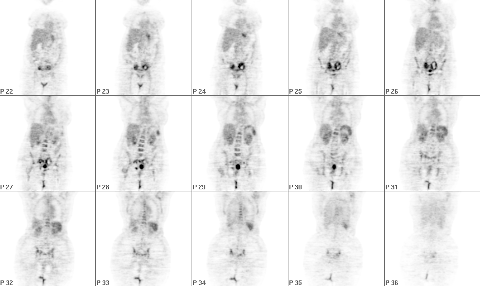
Whole-body coronal FDG-PET images are shown.
View main image(pt) in a separate image viewer
View second image(pt).
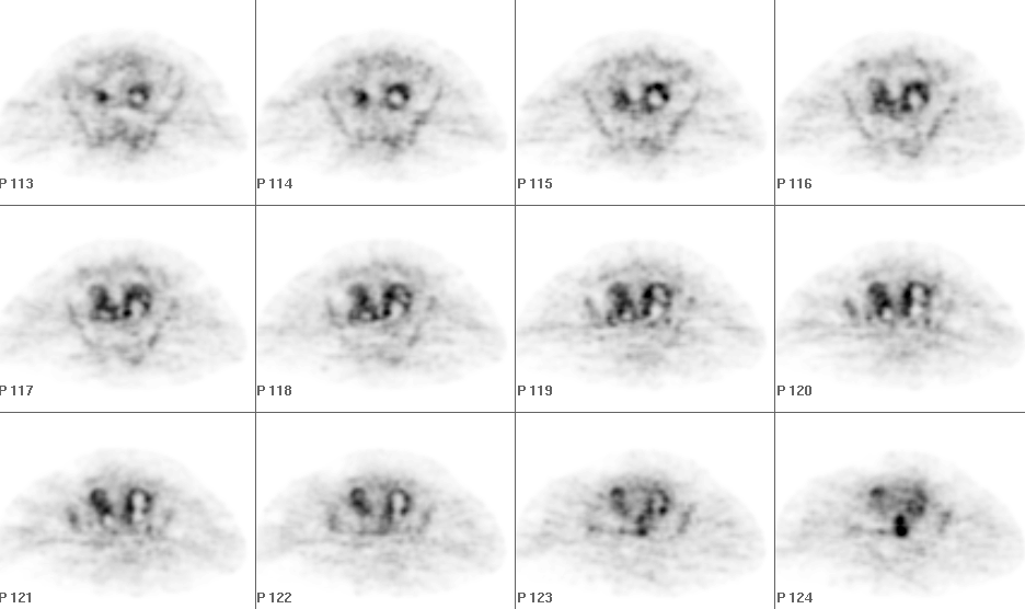
Axial FDG-PET images through the pelvis.
View third image(ct).
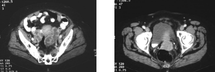
Axial CT images through the pelvis are shown.
View fourth image(ct).
Selected axial CT images of the pelvis.
Full history/Diagnosis is available below
Diagnosis: Pelvic inflammatory disease and cervical cancer.
Full history:
This is an 82 year old female with newly diagnosed invasive squamous cell carcinoma of the cervix being evaluated for staging.
Radiopharmaceutical:
15.0 mCi F-18 fluorodeoxyglucose (FDG) i.v.
Findings:
Whole-body PET images demonstrate an abnormal focus of very intense activity at the midline of the inferior pelvis. Additionally, in both adnexal regions, there are serpiginous foci of mild to moderate activity with nearly absent activity centrally. Note the physiologic activity in the gastic fundus and heterogeneously in the bone marrow. CT images demonstrate a soft tissue mass in the cervix at the midline and bilateral adnexal masses, which, upon closer inspection have the configuration of dilated fallopian tubes.
Discussion:
FDG-PET, while exquisitely sensitive for many malignancies is not entirely specific. In addition to neoplasm, any active infectious or inflammatory process can show increased FDG activity. In this case the patient's known primary cervical carcinoma was identified. The bilateral adnexal activity could represent metastatic spread, with central necrosis or tumor infiltrating the fallopian tube walls. However, given the milder FDG uptake compared with the primary cervical carcinoma, the delicate serpiginous distribution of activity, and the CT appearance, it was suggested that a process such as pelvic inflammatory disease was more likely.
Followup:
The patient progressed to a small bowel obstruction shortly after and was explored. At surgery she was found to have bilateral tubovarian abscesses without tumor infiltration. The primary cervical carcinoma was also identified.
ACR Codes and Keywords:
References and General Discussion of PET Tumor Imaging Studies (Anatomic field:Genitourinary System, Category:Inflammation,Infection)
Search for similar cases.
Edit this case
Add comments about this case
Return to the Teaching File home page.
Case number: pt030
Copyright by Wash U MO