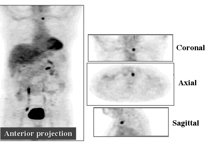
PET images
View main image(pt) in a separate image viewer
View second image(us). Thyroid ultrasonography.
Full history/Diagnosis is available below
Thyroid ultrasonography obtained following the PET study shows a 1.0 x 1.0 x 1.2 cm hypoechoic nodule in the inferior aspect of left thyroid lobe, correlating with the abnormality seen on FDG-PET.
The role of FDG-PET in thyroid carcinoma is unclear. Some have suggested it may find use in distinguishing follicular adenoma from follicular carcinoma (a difficult distinction to make with small tissue samples). I-123 imaging has been used for this purpose (normal or high uptake suggesting adenoma and low uptake suggesting tumor).
Several investigators report results indicating that FDG-PET may be useful for imaging and staging anaplastic thyroid tumors. Well-differentiated thyroid tumors that take up I-131 well commonly do not take up FDG well (95% per Feine, et al), while tumors that do not take up I-131 well appear to have high glucose metabolism and good FDG uptake.
References
Feine, U, et al. Fluorine-18-FDG and Iodine-131-Iodide uptake in thyroid cancer. J Nucl Med. 37, 1468-1471. 1996.
Gasparoni P. Potential role of fluorine-18-deoxyglucose (FDG) positron emission tomography (PET) in the staging of primitive and recurrent medullary thyroid carcinoma. J Endocrinol Invest. 20, 527-530. 1997.
Grünwald F, et al. Comparison of 18-FDG-PET with 131-iodide and 99m-Tc-sestamibi scintigraphy in differentiated thyroid cancer. Thyroid. 7, 327-335. 1997.
Rigo P, et al. Oncological applications of positron emission tomography with fluorine-18 fluorodeoxyglucose. Eur J Nucl Med. 23, 1641-1674. 1996.
Thyroidectomy was performed on 7/6/98, revealing papillary thyroid carcinoma, classical type. There was extension into the perithyroidal fat. Surgical margins were clear. One perithyroidal lymph nodes was positive for tumor. The patient subsequently underwent ablation of residual functioning thyroid tisse with I-131.
Two months later, CT of chest showed that the right lower lobe nodule was slowly enlarging and thus was suspicious for bronchogenic carcinoma. The nodule was resected on 8/10/98, revealing a necrotizing granuloma, with no evidence of acid-fast bacilli.
References and General Discussion of PET Tumor Imaging Studies (Anatomic field:Face, Mastoids, and Neck, Category:Neoplasm, Neoplastic-like condition)
Return to the Teaching File home page.