Case Author(s): John R. Leahy, M.D. and Barry A. Siegel, M.D. , 9/11/98 . Rating: #D3, #Q3
Diagnosis: Metastatic cervical carcinoma
Brief history:
38 year old woman with profuse vaginal bleeding.
Images:
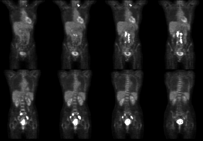
Contiguous coronal images
View main image(pt) in a separate image viewer
View second image(ct).
CT of the pelvis obtained several days prior to PET study
Full history/Diagnosis is available below
Diagnosis: Metastatic cervical carcinoma
Full history:
38 year old woman presented with vaginal bleeding. Cervical carcinoma
was diagnosed by biopsy. Subsequent CT scan revealed a large pelvic
mass and the patient was referred to Radiation Oncology for further
management.
Radiopharmaceutical:
15.0 mCi F-18 fluorodeoxyglucose i.v.
Findings:
Several foci of increased F-18 fluorodeoxyglucose (FDG) accumulation are
seen. This includes a large, heterogeneous pelvic mass, which correlates
to the cervical mass noted on CT, and is consistent with the patient's
primary tumor. There are also several foci of intense FDG
accumulation along the courses of both iliac nodal chains with
extension along the course of the periaortic nodal chains. An
additional focus of intense activity is seen in the left supraclavicular
region, likely representing a lymph node metastasis. The results of PET
are therefore consistent with stage IV cervical cancer. The degree of
lymphadenopathy demonstrated on this PET study is more extensive than
that seen on CT. No hydronephrosis is appreciated.
Discussion:
Cervical carcinoma metastasizes from the primary site in a predictable
pattern. Disease extends through pelvic lymph nodes, iliac chains,
and along the aorta, before finally to spreading to extranodal sites
such as lung or liver. The presence of lymph node metastases does not
alter the FIGO clinical stage, but indicates a worse prognosois and can
influence the choice of therapy. Detection of lymph node involvement by
CT is limited by size criteria, having a sensitivity of 34% and
specificity of 96%. PET depends on metabolic rather than size criteria
for detection of nodal involvement. Preliminary data show that PET can
detect metastatic disease, both locoregional and distant, with greater
sensitivity than conventional imaging studies, as is evidenced in this
study. PET also shows utility in assessment of recurrent disease.
References:
Grigsby PW, Dehdashti F, Siegel BA. FDG-PET evaluation of carcinoma of
the cervix. Clin Positron Imaging 1999; 2:105-9.
Rose PG, Adler LP, Rodriguez M, Faulhaber PF, Abdul-Karim FW, Miraldi
F. Positron emission tomography for evaluating para-aortic nodal
metastasis in locally advanced cervical cancer before surgical staging:
a surgicopathologic study. J Clin Oncol 1999; 17:41-5.
Sugawara Y, Eisbruch A, Kosuda S, Recker BE, Kison PV, Wahl RL.
Evaluation of FDG PET in patients with cervical cancer. J Nucl Med
1999; 40:1125-31.
Followup:
The woman received implant radiation and chemotherapy for the primary
tumor. She also underwent external beam radiation to the
abdominopelvic and supraclavicular lymph node metastases detected by
PET. She returned on 8/11/98 for interval evaluation of her disease.
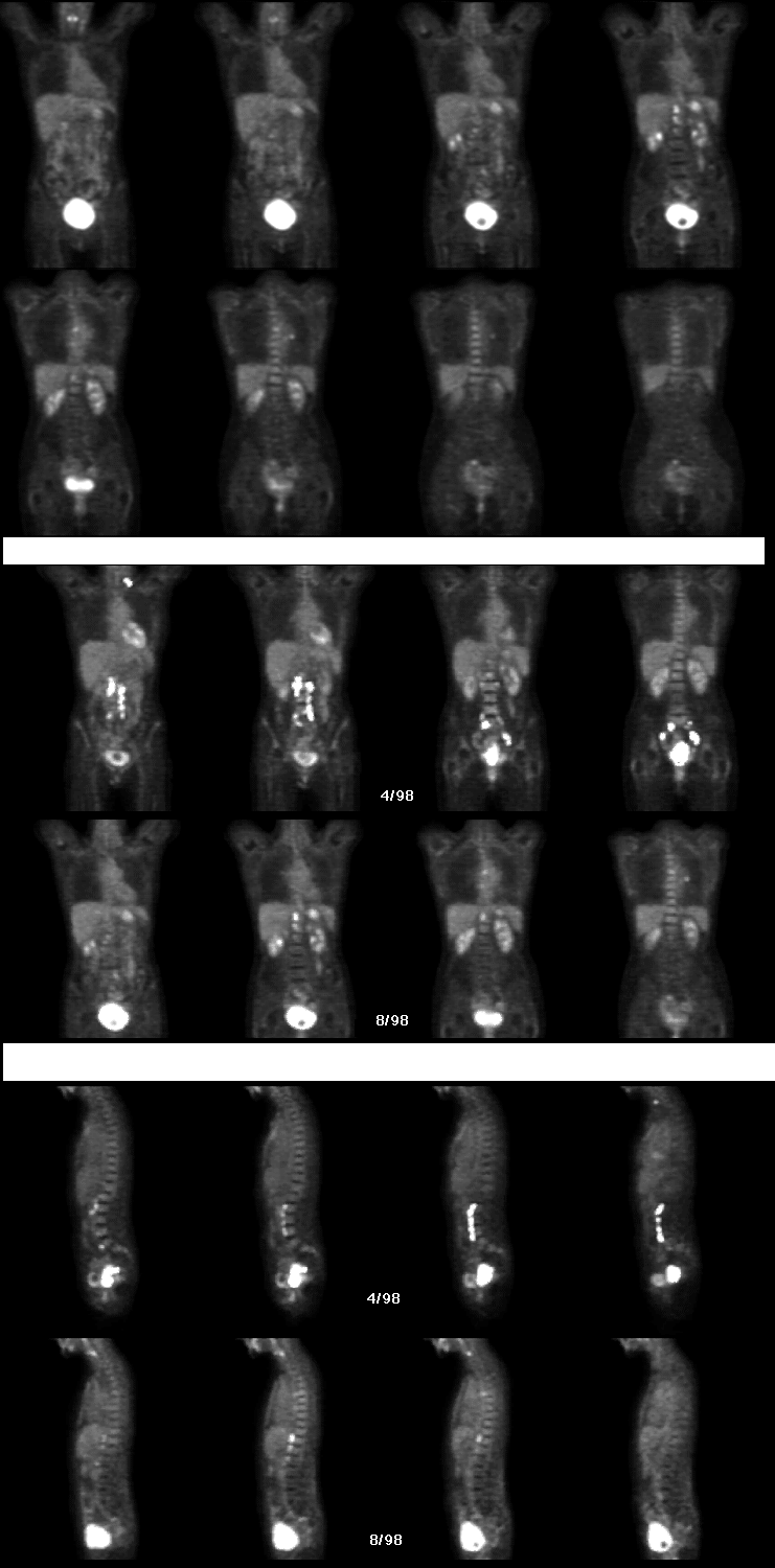
Findings:
The first set of images--coronal images obtained on 8/11/98--shows resolution
of activity in the primary tumor and the original lymph node metastases
(because of a malfunctioning Foley catheter, there is a large amount of urine in the
bladder). There are three new foci of abnormal FDG accumulation in the mid
abdominal periaortic lymph nodes, as well as two new foci in the
posterior mediastinum and left pulmonary hilum.
The second and third sets--comparison coronal and sagittal images--more
clearly show the interval changes from April to August. There is
decreased marrow activity extending from the sacrum to the L1
level. This suggests fatty marrow replacement, and corresponds to the
radiation port used to treat the original lymph node metastases. Note
that metastatic disease has now developed in the nodal chain just above
the radiation port.
Given the rapid development of the new foci, they likely arose from
micrometastases present, but not detectected on the first PET
study. These foci were not covered in the radiation port.
View followup image(pt).
Three sets of images: 1. Coronal images from 8/11/98. 2. Comparison of
coronal images from 4/98 and 8/98. 3. Comparison of sagittal images from
4/98 and 8/98
ACR Codes and Keywords:
References and General Discussion of PET Tumor Imaging Studies (Anatomic field:Genitourinary System, Category:Neoplasm, Neoplastic-like condition)
Search for similar cases.
Edit this case
Add comments about this case
Read comments about this case
Return to the Teaching File home page.
Case number: pt020
Copyright by Wash U MO