Case Author(s): michael quinn, md and farrokh dehdashti, md , 4-10-98 . Rating: #D2, #Q4
Diagnosis: sarcoid and colon cancer metastases
Brief history:
47 yo male with colon carcinoma
Images:
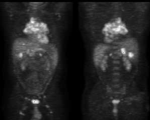
anterior and posterior images are shown
View main image(pt) in a separate image viewer
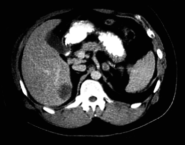
View second image(ct).
abdominal ct image
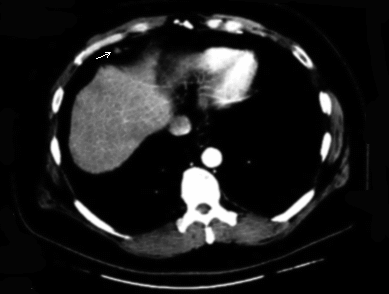
View third image(ct).
abdominal ct image
Full history/Diagnosis is available below
Diagnosis: sarcoid and colon cancer metastases
Full history:
47 yo old male with prior colon cancer who had two suspicious liver
lesions on CT scan. The PET study was performed to evaluate these
lesions as well as other possible sites of disease. Other pertinent
clinical history was not provided at the time of the study.
Radiopharmaceutical:
PET-FDG
Findings:
There are multiple foci of abnormal activity throughout the body. The
known liver abnormalities are metabolically active. Additionally,
marked perihilar and mediastinal activity is present. Smaller
foci are seen in the porta hepatis, retroperitoneum, inguinal regions,
right axillary region, and right supraclavicular region.
Discussion:
Given the patients prior history of colon carcinoma, the abnormalities
in the liver are suspicious for metastases as they display marked
glucose metabolism. However, the remainder of the abnormal foci
distributed throughout the chest, abdomen, and pelvis would be an
unusual appearance of colon cancer metastases. A better explanation
would be that of a more systemic process. After contacting the
referring physician, it was found that the patient also carried an
underlying diagnosis of sarcoidosis.
Sarcoidosis is a chronic granulomatous inflammatory disease that is of
unknown origin. While it commonly affects the lungs, nearly any
tissue in the body may be affected. The initial inflammatory infiltrate
consists of mononuclear cells, with subsequent formation
of granulomas in the affected tissues. These granulomas may go on to
resolve or lead to fibrosis. During the inflammatory phase there is
increased glycolysis related to the metabolic activity of the cellular
infiltrate. This is reflected in the increased activity seen in these
same regions on PET-FDG studies during this phase. However, this is a
nonspecific finding which may also be seen at other sites of
inflammation (i.e. infection) as well as in malignancies. FDG uptake has been seen in vitro to be accumulated by leukocytes, lymphocytes and macrophages and FDG uptake is seen in vivo at sites of infection. Therefore, a
concern exists for false positive interpretation for metastases in
patients with coexisting malignancy and a systemic metabolic disease
such as sarcoid. Use of other imaging modalities to discern the two as
well as knowledge of likely routes of metastatic spread help to decrease
the risk of overcalling the extent of tumor spread.
References:
1. Lewis and Salama, Uptake of Fluorine-18- Fluorodeoxyglucose
in Sarcoidosis. J Nucl Med 1994; 35:1647-1649
1. Alavi et al (Editorial) Is There A Role For PET-FDG
Imaging In The Management Of Patients With Sarcoidosis? J Nucl Med 1994;
35: 1650-1652
Differential Diagnosis List
lymphoma
ACR Codes and Keywords:
References and General Discussion of PET Tumor Imaging Studies (Anatomic field:Vascular and Lymphatic Systems, Category:Inflammation,Infection)
Search for similar cases.
Edit this case
Add comments about this case
Return to the Teaching File home page.
Case number: pt019
Copyright by Wash U MO