Case Author(s): Michael Quinn, M.D. and Barry A. Siegel, M.D. , 09/11/97 . Rating: #D3, #Q4
Diagnosis: Attenuation-Correction Artifact on FDG-PET
Brief history:
This 47-year-old woman has a history of treated
dermatofibrosarcoma and now has a lung nodule seen on CT.
This study was requested to evaluate for metastatic disease.
Images:
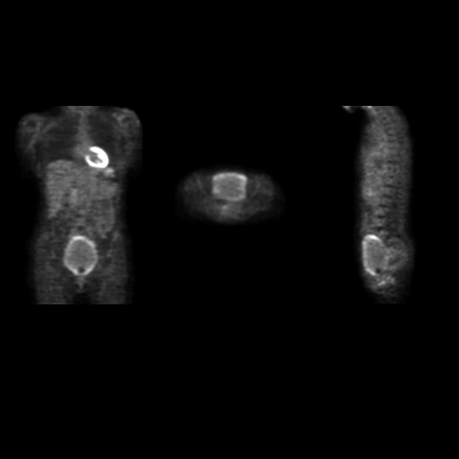
Coronal, axial, and sagittal emission images from a whole-body FDG-PET study
are shown.
View main image(pt) in a separate image viewer
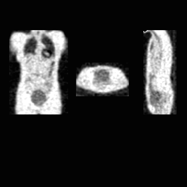
View second image(pt).
Coronal, axial, and sagittal images transmission images are shown.
Full history/Diagnosis is available below
Diagnosis: Attenuation-Correction Artifact on FDG-PET
Full history:
This 47-year-old woman has a history of a left shoulder
dermatofibrosarcoma treated by resection. Radiographs and CT of the chest
revealed a nodule which was considered suspicious for a metastatic focus
in the right upper lobe. An FDG-PET study was ordered to evaluate
for metabolic activity in this nodule.
Radiopharmaceutical:
F-18 fluorodeoxyglucose (FDG)
Findings:
No abnormal activity was seen either in the site of the patient's
prior primary tumor or at the site of the lung nodule. This
suggests that the lung nodule is benign. However, the emission images
are remarkable for a rim of increased activity along the periphery of the
bladder. There are no other abnormalities.
Discussion:
At Mallinckrodt Institute of Radiology, all PET tumor studies are reconstructed with use of
a computer algorithm that takes into account the attenuation of emitted
photons by the patient's tissues. This "attenuation correction" is
achieved by obtaining initial transmission images with the patient
positioned between a rotating germanium source and the camera detectors.
Regions of low photon attenuation (lungs) result in more activity from
the source being detected by the scanner. Conversely, regions of increased
attenuation (abdomen) result in fewer counts detected from the
transmission source. These data are used to create an attenuation "map"
corresponding to each cross-section through the patient. The subsequent
emission images then use this map to take into account regional photon
attenuation of administered radiopharmaceutical by various soft tissues.
However, with the technique we employ, the FDG is administered before
the patient is put into the scanner, and is thus "on board" at the time
of transmission imaging. Regions with marked radiopharmaceutical
accumulation, such as the myocardium and bladder, have resultant large numbers of photons detected
on transmission scan. The detector cannot differentiate photons emitted
by the FDG from those arising from the transmission source. These areas
are thus interpreted as having little photon attenuation, because so many counts are
detected. This causes subsequent under-correction of these same regions
on the corresponding emission images. Such under-correction is usually
limted to large, very intense regions like the bladder, where
interpretation is rarely an issue.
We routinely place a Foley catheter for FDG-PET tumor imaging studies, and
also administer intravenous fluids and furosemide to minimize the
activity in the pelvicalyceal systems of the kidneys and in the urinary
bladder. However, in this patient, there was a significant increase in
bladder volume during the interval between transmission and emission
imaging, presumably because the Foley catheter was not draining properly.
On the emission images, the central bladder is under-corrected, as
expected, whereas a surrounding rim of urine accumulating in the
interval between scans, is accurately corrected for attenuation. This
resulted in the erroneous appearance of an "abnormal" rim of activity
at the periphery of the bladder.
Although unlikely, potential non-artifactual causes of the FDG distribution
pattern seen in this patient would include cystitis and a diffuse mucosal or mural
neoplasm of the bladder.
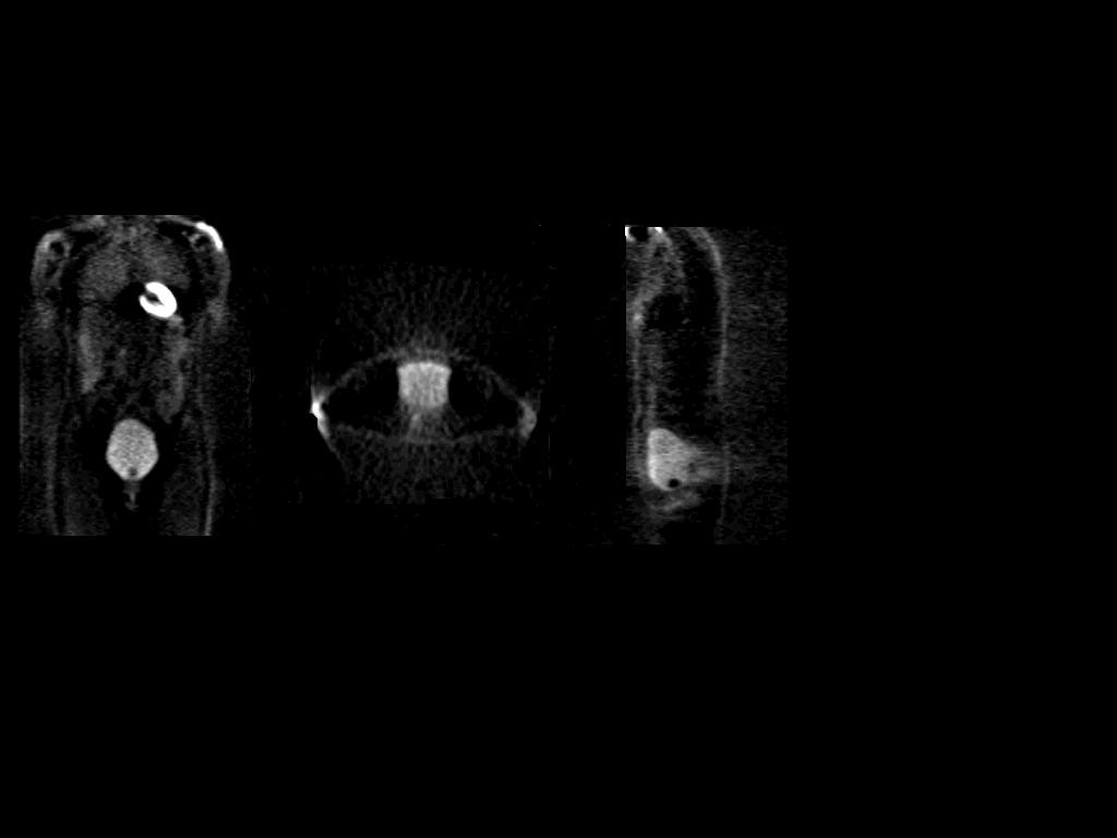
Followup:
To confirm the artifactual nature of the finding, images were
reconstructed from the emission data without use of attenuation
correction. These showed more uniform activity in the enlarged bladder.
The slightly greater peripheral than central activity is related to the
the effect of attenuation of photons arising deeper within the body.
View followup image(pt).
Coronal, axial, and sagittal emission images (without attenuation correction)
from a whole-body FDG-PET study
are shown
Major teaching point(s):
For accurate attenuation correction there must be no change in the
object between emission and transmission images. The most likely source
of artifacts due to such changes is patient motion, but changes in
internal organ configuration (e.g., in distribution of bowel gas or,
as in this case, in bladder volume) can also cause artifacts.
Differential Diagnosis List
As noted above, cystitis and a diffuse mucosal or mural
neoplasm of the bladder could produce similar findings.
ACR Codes and Keywords:
References and General Discussion of PET Tumor Imaging Studies (Anatomic field:Lung, Mediastinum, and Pleura, Category:Other(Artifact))
Search for similar cases.
Edit this case
Add comments about this case
Return to the Teaching File home page.
Case number: pt016
Copyright by Wash U MO