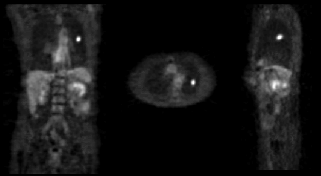
CORONAL, AXIAL, AND SAGITTAL SLICES
View main image(pt) in a separate image viewer
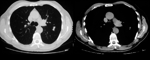
View second image(ct). CT IMAGES THRU THE UPPER LUNG FIELDS (MEDIASTINAL AND LUNG WINDOWS)
Full history/Diagnosis is available below
CT of the chest shows a 2 x 3 x 1.5 cm left upper lobe nodule with straight margins (atypical for malignancy), but no definite benign-appearing calcification. Additional axial slices (not included) showed calcified mediastinal lymph nodes consistent with old granulomatous disease.
PET is useful for the further characterization of indeterminant lung nodules in that it helps stratify patients into those that need further work-up and biopsy and those who can be managed with watchful waiting. Ideally, the nodule should be greater than 8-10 mm in size due to limitations in PET resolution.
Nodules showing relative increased tracer uptake (ie. increased metabolic activity) are highly sensitive (93-100%) and slightly less specific (78-90%) for malignancy. Semi-quantitative analysis using the standardized uptake value (SUV) is an objective measure of the lesion's metabolic activity when compared with the average concentration of FDG within the body. When evaluating lung nodules, any SUV greater than 2.5 is considered positive and malignancy is found in > 83%. If the nodule has an SUV < 2.5, then malignancy is seen in < 5%.
As in this case, most false positives for malignancy are due to active granulomatous disease.
In the evaluation of indeterminant lung nodules, many patients with benign disease can be spared the morbidity of biopsy if initial work-up includes a negative PET scan.
If the nodule is less than 8-10 mm in size, then all bets are off.
Granulomatous disease
References and General Discussion of PET Tumor Imaging Studies (Anatomic field:Lung, Mediastinum, and Pleura, Category:Inflammation,Infection)
Return to the Teaching File home page.