Case Author(s): Michael Quinn, MD Farrokh Dehdashti, MD , 07/18/97 . Rating: #D2, #Q3
Diagnosis: Mesothelioma
Brief history:
65 year old male with right upper extremity pain.
Images:

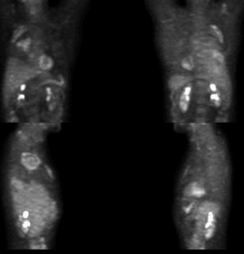
Reprojection images in anterior, posterior, and lateral position.
View main image(pt) in a separate image viewer
View second image(pt).
Cross sectional images as labeled
Full history/Diagnosis is available below
Diagnosis: Mesothelioma
Full history:
65 year old male who initially presented with right upper extremity
pain. Work-up revealed a mass in the peripheral right lung apex.
There was associated rib destruction and pleural thickening. Biopsy
was non diagnostic.
Radiopharmaceutical:
FDG
Findings:
The PET images are markedly abnormal. There is increased activity in
the right lung apex corresponding to the mass seen on CT in this
location.The peripheral location of the mass is again noted. Of
interest is the lenticular shape of the mass, with convex margins on
both sides. Accumulation of activity is increased laterally compared
with medially, and these is a paucity of activity centrally. There is
patchy intense accumulation in the anterosuperior right pleura and
posterobasal right pleura which corresponds to the regions of pleural
thickening seen on CT. Moderate activity in the right anterior sulcus
is present, as is a small focus in the right hilum. There is a focus of activity
in the right abdomen.
Discussion:
Following the patient's initial CT, a needle biopsy of the right
apical mass was performed. Unfortunately, this specimen was deemed
non-diagnostic by pathology. The PET study clearly shows that the
mass has a photopenic center. It was surmised
that the biopsy was taken from this area and may have sampled the
necrotic/fibrotic center of a mass. As a tissue diagnosis was necessary
to determine treatment in this patient, a second biopsy was planned.
The PET study was used to localize another site of biopsy in the chest
to avoid another non-diagnostic study. One of the peripheral regions
of activity corresponding to a pleural based lesion on CT was chosen.
The subsequent biopsy of this region yielded a diagnosis of
mesothelioma.
Anatomical imaging modalities are limited in differentiating benign from
malignant pleural abnormalities. The utility of FDG-PET in this clinical
setting has been assessed in a limited fashion. Bury et al have
demonstrated that FDG-PET has a sensitivity of 94% and specificity of 78%
for differentiating benign from malignant pleural abnormalities.
Similar results have been reported by Lowe et al (sensitivity of 94% and
specificity of 67%). Malignant pleural disease typically exhibits
moderate to marked FDG accumulation. A lack of FDG uptake within the
pleural lesion has a high negative predictive value for absence of
malignant disease. In both reports, false-positive results were seen
in patients with infectious/inflammatory pleural disease. As shown
in the above case, FDG-PET can be used for biospy guidance for
histological confirmation.
References:
1) Knight SB, Delbeke D, Stewart JR, Sandler MP. Evaluation of
pulmonary lesions with FDG-PET. Comparison of findings in patients
with and without a history of prior malignancy.
Chest 1996 Apr; 109(4):982-8.
2) Lowe VJ, Patz E, Harris L, Hoffman JM, Hanson M, Goodman P,
Coleman RE. FDG-PET evaluation of pleural abnormalities.
J Nucl Med 1994; 35:228P.
View followup image(ct).
Transaxial CT slice, mid thorax
Differential Diagnosis List
Tumor
ACR Codes and Keywords:
- General ACR code: 63
- Lung, Mediastinum, and Pleura:
6.3254 "Pleural include: mesothelioma"
References and General Discussion of PET Tumor Imaging Studies (Anatomic field:Lung, Mediastinum, and Pleura, Category:Neoplasm, Neoplastic-like condition)
Search for similar cases.
Edit this case
Add comments about this case
Return to the Teaching File home page.
Case number: pt014
Copyright by Wash U MO

