Case Author(s): Scott Winner, M.D. and Farrokh Dehdashti M.D. , 11/01/96 . Rating: #D4, #Q3
Diagnosis: tortuous, ectatic ureter
Brief history:
63-year old man with a
history of rectal adenocarcinoma
Images:

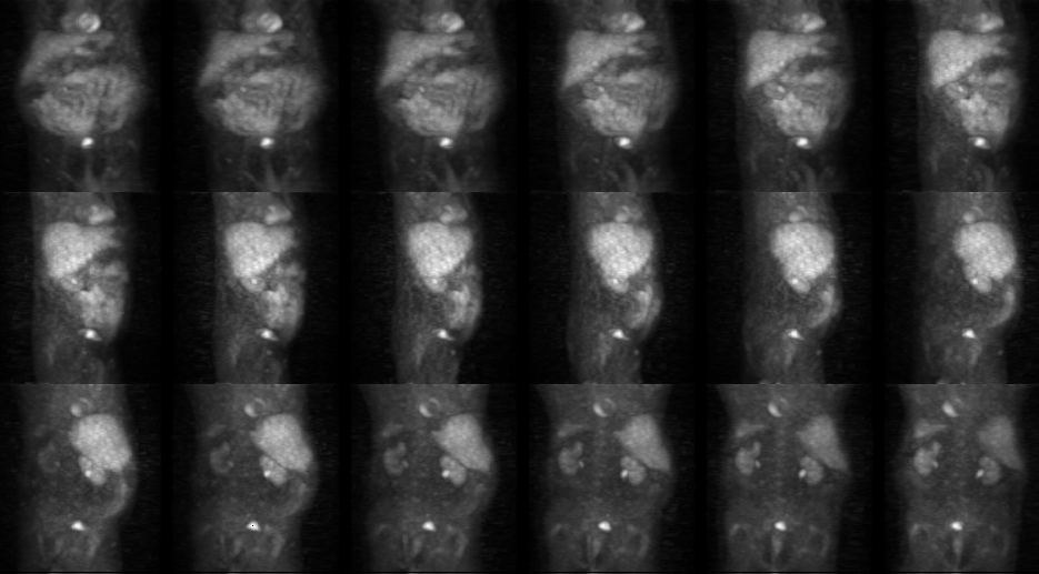
PET images progressing from anterior to posterior.
View main image(pt) in a separate image viewer
View second image(ct).
CT of the pelvis without contrast
View third image(ct).
CT of the pelvis with contrast
Full history/Diagnosis is available below
Diagnosis: tortuous, ectatic ureter
Full history:
The patient has a history of
rectal adenocarcinoma, which was treated with
preoperative radiotherapy. The patientıs
carcinoma was resected on 9-9-95 with a tumor
stage of T3, N0, M0. A CT scan done without
contrast on 9-17-96 demonstrated a 1.2 cm
right perirectal lymph node. The patient
presented for further evaluation with positron
emission tomography on 10-1-96.
Radiopharmaceutical:
14.7 mCi F-18
fluorodeoxyglucose
Findings:
There is a focus of intense
FDG accumulation in the pelvis which
corresponds in location with the right
perirectal lymph node seen on the CT of 9-17-
96. This is suspicious for metastatic disease.
Discussion:
The non-contrast pelvic CT
of 9-17-96 and the PET study of 10-1-96 were
false-positive studies. Further evaluation with a
contrast enhanced CT on 10-30-96
demonstrated that the suspected 1.2 cm right
perirectal lymph node was in fact a slightly
dilated tortuous right ureter. The activity seen
at this location on the PET study represented
normal FDG pooling within the ureter. FDG is cleared
from the circulation by renal excretion (the
mechanism of renal clearence of FDG is not well
understood). Activity within the ureters and
bladder is normally seen and can sometimes mimic pathology.
Followup:
None.
Major teaching point(s):
A careful correlation with cross-sectional imaging is essential to avoid misinterpretation of the PET images.
ACR Codes and Keywords:
- General ACR code: 81
- Genitourinary System:
8.13 "NORMAL VARIANT"
References and General Discussion of PET Tumor Imaging Studies (Anatomic field:Genitourinary System, Category:Normal, Technique, Congenital Anomaly)
Search for similar cases.
Edit this case
Add comments about this case
Read comments about this case
Return to the Teaching File home page.
Case number: pt013
Copyright by Wash U MO

