Case Author(s): Ed Grishaw, M.D. and Farrokh Dehdashti, M.D. , 08/02/96 . Rating: #D3, #Q4
Diagnosis: Esophageal carcinoma metastatic to the liver and lymph nodes.
Brief history:
52-year old man
with upper gastrointestinal bleeding.
Images:

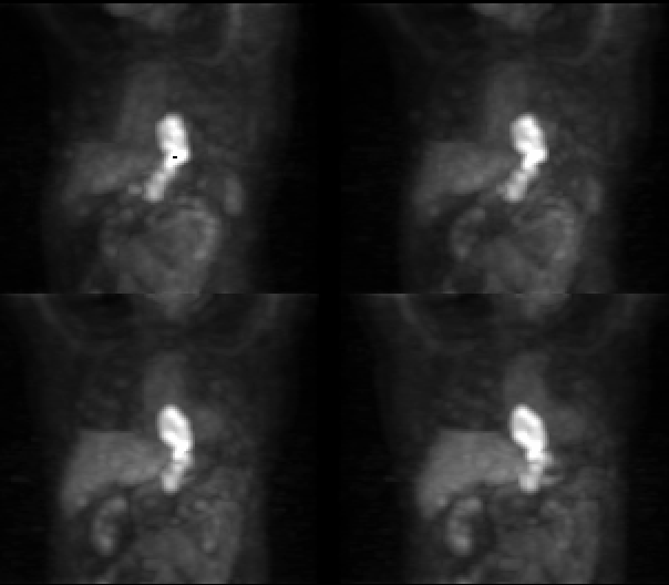
Volume rendered images from anterior and anterior-oblique views.
View main image(pt) in a separate image viewer
View second image(pt).
Coronal, transaxial, and sagittal slices.
View third image(ct).
CT images at several levels
Full history/Diagnosis is available below
Diagnosis: Esophageal carcinoma metastatic to the liver and lymph nodes.
Full history:
52-year old man
with upper gastrointestinal bleeding.
Subsequent CT scan of the chest and
upper abdomen demonstrated an
ulcerated esophageal mass extending
from the mid thoracic esophagus to the
gastroesophageal junction. Biopsy of
the mass revealed a mildly
differentiated adenocarcinoma.
Radiopharmaceutical:
16.4 mCi
F-18 fluorodeoxyglucose i.v.
Findings:
The PET images
demonstrate marked heterogeneously
increased FDG accumulation beginning
just distal to the hila extending to the
gastroesophageal junction There is
possible involvement of the gastric
cardia . Focal, nodular increased FDG
accumulation is seen in the region of
the gastrohepatic ligament and celiac
axis corresponding to the adenopathy
identified on computed tomography .
In addition, a small focus of
hypermetabolism is identified in the
right lobe of the liver (segment 5),
compatible with a metastatic lesion.
No corresponding abnormality can be
identified on computed tomography.
Discussion:
Malignant esophageal
lesions outnumber benign tumors by
more than four to one. The majority of
malignant esophageal tumors are
squamous cell carcinomas; occasional
examples of primary esophageal
adenocarcinoma, carcinosarcoma,
lymphoma, sarcoma, and melanoma.
have been described At the time of
diagnosis, neoplastic burden is usually
advanced with the overall five-year
survival rate between 4% and 10%
despite more aggressive surgical and
radiation therapies. Direct contiguous
and lymphatic spread can be rapid
because of the lack of an esophageal
serosa.
References:
Moss AA,
Gamsu G, Genant HK. Computed
tomography of the body with magnetic
resonance imaging: Volume 3,
Abdomen and Pelvis. W.B. Saunders
Co., 1992; pp 649-659.
Flanagan FL, Dehdashti F, Siegel BA, Trask DD, Sundaresan SR,
Patterson GA, Cooper JD. Staging of esophageal cancer with FDG-PET.
AJR 1997: 168:417-424.
Major teaching point(s):
This case
illustrates the utility of PET in the
pretreatment planning of esophageal
carcinoma. Its utility not only lies in its
ability to detect the extent of
theprimary lesion, but also areas of
metastatic disease, which in some cases
may be subradiographic.
ACR Codes and Keywords:
References and General Discussion of PET Tumor Imaging Studies (Anatomic field:Gasterointestinal System, Category:Neoplasm, Neoplastic-like condition)
Search for similar cases.
Edit this case
Add comments about this case
Return to the Teaching File home page.
Case number: pt012
Copyright by Wash U MO

