Case Author(s): Charles Pringle, M.D., and Farrokh Dehdashti, M.D. , 06/03/96 . Rating: #D4, #Q4
Diagnosis: Recurrent(residual) left frontal astrocytoma.
Brief history:
History of brain tumor with
recent syncopal episode
Images:

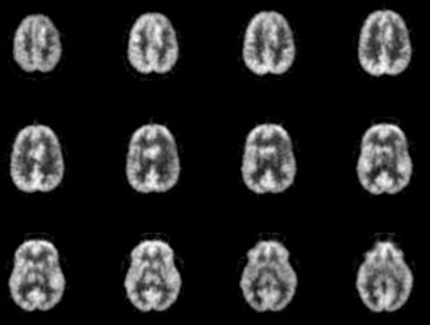
Axial images
View main image(pt) in a separate image viewer
View second image(pt).
Coronal P.E.T. images
View third image(mr).
Axial T2 images
View fourth image(mr).
Coronal T1 with contrast images
Full history/Diagnosis is available below
Diagnosis: Recurrent(residual) left frontal astrocytoma.
Full history:
31-year old man with history of
grade II astrocytoma of the left frontal lobe, status
post resection four years previously and also external
beam radiation therapy. The patient had done well
until recently when he experienced a syncopal episode
at work.
Radiopharmaceutical:
10.8 mCi F-18
fluorodeoxyglucose i.v.
Findings:
There is intense abnormal FDG
accumulation in the left frontal lobe, which extends
across the mid line in the region of the corpus
callosum. This pattern does correspond to the
abnormal signal on the MRI examination in the same
region. The MRI also demonstrated an area of
abnormal signal in the left parietal lobe with
peripheral enhancement. No corresponding PET
abnormality was noted. It has been shown that in brain tumors following therapy, FDG uptake into a contrast-enchancing lesion suggests the presence of viable tumor, while absent FDG uptake suggests necrosis. FDG PET is very useful in differentiating residual/recurrent tumor from post-therapeutic changes as post-therapy MRI is unable to differentiate tumor from benign changes due to therapy.
Discussion:
This study demonstrates the
utility of PET imaging in the evaluation of sites of
previous tumor within the brain which could
represent recurrent or residual tumor vs
postoperative changes.
ACR Codes and Keywords:
References and General Discussion of PET Tumor Imaging Studies (Anatomic field:Skull and Contents, Category:Neoplasm, Neoplastic-like condition)
Search for similar cases.
Edit this case
Add comments about this case
Read comments about this case
Return to the Teaching File home page.
Case number: pt010
Copyright by Wash U MO

