Case Author(s): Gregg D. Schubach, M.D., Philip Moyers, M.D., and Farrokh Dehdashti, M.D. , . Rating: #D3, #Q3
Diagnosis: Primary Pancreatic Carcinoma with metastatic lung carcinoma to left adrenal
Brief history:
58-year old man status post
surgically resected right upper lobe adenocarcinoma
has two abnormalities on a recent CT study of the
abdomen.
Images:

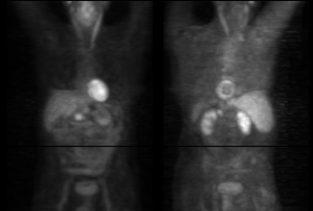
Anterior and slight RPO projections from PET study
View main image(pt) in a separate image viewer
View second image(ct).
Axial CT image from level of pancreas
View third image(ct).
Axial CT image from level of adrenal
Full history/Diagnosis is available below
Diagnosis: Primary Pancreatic Carcinoma with metastatic lung carcinoma to left adrenal
Full history:
58-year old man status post
surgically resected right upper lobe adenocarcinoma
and radiation therapy has a pancreatic head mass and
left adrenal mass on a recent CT study of the
abdomen.
Radiopharmaceutical:
F-18
Fluorodeoxyglucose
Findings:
FDG whole body PET imaging
demonstrates markedly increased FDG accumulation
in the pancreatic head (on the anterior image) and in
the left adrenal gland (on the posterior image). The
CT images demonstrate a soft tissue mass in the head
of the head of the pancreas and a nodule in the left
adrenal gland.
Discussion:
Positron emission tomography
(PET) is a unique imaging modality that obtains
functional imaging data from the human body. The
count density in reconstructed PET images, unlike
other radionuclide imaging techniques, is directly
proportional to the local radioactivity concentration.
Therefore, tracer uptake may be absolutely
quantified. PET plays a complementary
role to other imaging modalities, such as magnetic
resonance imaging (MRI) or computed tomography
(CT), which mainly provide anatomic rather than
functional information. Functional changes may
proceed anatomic changes in many disorders, such as
Alzheimeršs disease, Parkinsonšs disease,
Huntingtonšs disease, neurodevelopmental disorders,
psychiatric disorders, epilepsy, cardiomyopathies,
coronary artery disease, and a whole host of
malignancies.
The clinical oncologic applications of PET, while not
as well developed as cardiac and neurologic
applications, appear very promising and most
investigators foresee several major clinical
applications within the next five years.
Fluorodeoxyglucose (FDG)-PET studies have high
sensitivity, specificity, and accuracy for evaluating
radiologically indeterminate solitary pulmonary
nodules and accuracy in staging lung tumors.
Differentiating residual post-surgical scar from
persistent tumor is a common clinical problem, which
PET may resolve. Gupta, et al. and Knapp, et al., in
separate studies, demonstrate 100% specificity in
their studies of 19 and 18 patients, respectively.
Potential indications for PET in clinical oncology during initial
evaluation include: 1) differentiating benign from
malignant lesion; 2) determining degree of
malignancy; 3) localizing site for biopsy; 4) staging;
and 5) assessing prognosis. Following therapy, the
clinical indications for PET include: 1) documenting
recurrence; 2) assessing persistence; 3) determining
effectiveness of treatment; and 4) monitoring
progression.
References: Coleman, R.E., editor, The Radiologic Clinics of
North America, July 1993 31:4 2) Sandler, M.P., et al.
Diagnostic Nuclear Medicine, 3rd edition, 1996.
Williams and Wilkins
Followup:
Biopsy of the left adrenal nodule
under CT guidance revealed adenocarcinoma (most
compatible with pancreatic adenocarcinoma primary). The metastatic adrenal
adenocarcinoma is likely from the bronchogenic carcinoma in this patient, although it could be from pancreatic carcinoma.
Differential Diagnosis List
Increased FDG uptake is not as specific for malignancy and can be seen in pheochromocytomas.
ACR Codes and Keywords:
References and General Discussion of PET Tumor Imaging Studies (Anatomic field:Gasterointestinal System, Category:Neoplasm, Neoplastic-like condition)
Search for similar cases.
Edit this case
Add comments about this case
Read comments about this case
Return to the Teaching File home page.
Case number: pt009
Copyright by Wash U MO

