Case Author(s): Charles Pringle, M.D. and Farrokh Dehdashti, M.D. , 01/11/96 . Rating: #D3, #Q3
Diagnosis: Early fibrosing mediastinitis
Brief history:
Dysphagia
Images:

Anterior, posterior, and lateral images
View main image(pt) in a separate image viewer
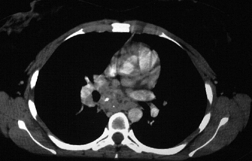
View second image(ct).
axial enhanced image through mediastinum
Full history/Diagnosis is available below
Diagnosis: Early fibrosing mediastinitis
Full history:
26-year old woman with two-
week history of dysphagia. A recent CT scan showed
a soft tissue mass in the mid esophagus and barium
swallow showed an extrinsic compression upon the
mid esophagus, with a smooth appearing mucosa of
the esophagus in that area.
Radiopharmaceutical:
15.8 mCi F-18
fluorodeoxyglucose (FDG) i.v.
Findings:
There is focally abnormal intense
FDG accumulation in the region of the mid esophagus
with an irregular appearance along the inferior and
right lateral margins. This area does correspond to a
soft tissue mass identified on CT scan, which could be
due to a malignant process or an active inflammatory
disease.
Discussion:
The CT scan findings of a soft
tissue mass with some small calcifications and the
barium swallow demonstrating extrinsic pressure
upon the esophagus were most consistent with a
granulomatous process and most likely represent
early fibrosing mediastinitis.
References:
1) J Nucl Med 1992;33:1972-1980
2) J Nucl Med 1975;15:352-355
Followup:
Patient was treated for presumed
fibrosing mediastinitis.
Major teaching point(s):
The appearance of increased
FDG accumulation in the region of the soft tissue
mass is consistent with increased metabolism and can
be present in active granulomatous processes in
addition to neoplasm. The type of inflammatory
responses are important in determining the degree of
FDG uptake. Tuberculosis, fungal infections, and
abscess have all been associated with increased FDG
uptake. These infections are characterized by cellular
infiltrates, granuloma formation, and macrophage
proliferation. Activated inflammatory cells have a
markedly increased glycosis and the hexose
monophosphate shunt is stimulated by phagocytosis
with increases of 20-30 times baseline being common
in these cells.
Differential Diagnosis List
Active
granulomatous disease, malignant tumor.
ACR Codes and Keywords:
References and General Discussion of PET Tumor Imaging Studies (Anatomic field:Lung, Mediastinum, and Pleura, Category:Inflammation,Infection)
Search for similar cases.
Edit this case
Add comments about this case
Read comments about this case
Return to the Teaching File home page.
Case number: pt008
Copyright by Wash U MO