Case Author(s): J. Philip Moyers, M.D. and Farrokh Dehdashti, M.D. , 12/5/95 . Rating: #D3, #Q3
Diagnosis: Metastatic uterine leiomyosarcoma
Brief history:
Patient is status post
hysterectomy
Images:

Anterior, posterior and bilateral lateral views from a whole body PET study
View main image(pt) in a separate image viewer
View second image(xr).
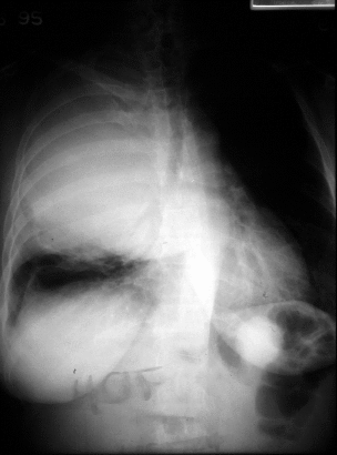
PA chest radiograph
View third image(ct).
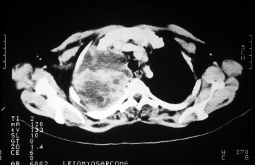
Axial Section from chest CT, soft tissue windows
View fourth image(ct).
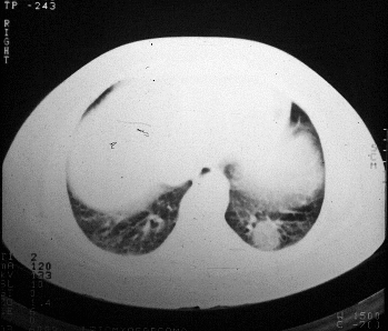
Axial section from chest CT, lung windows
Full history/Diagnosis is available below
Diagnosis: Metastatic uterine leiomyosarcoma
Full history:
40-year old woman with a uterine
leiomyosarcoma diagnosed in August 1993, now status
post hysterectomy and radiotherapy. Over the last
two months, the patient has had cough and daily
fever. Computed tomographic examination of the
chest after chest radiograph revealed a large right
upper hemithorax mass and left lower lobe mass. The
right-sided mass was biopsied under CT guidance,
revealing metastatic leiomyosarcoma with histologic
features similar to the primary tumor. This
examination is requested to evaluate extent of
metastatic disease prior to debulking surgery due to
the patientąs respiratory compromise.
Radiopharmaceutical:
15.8 mCi F-18
fluorodeoxyglucose (FDG) i.v.
Findings:
Abnormal foci of increased activity
are demonstrated in the right upper hemithorax as
well as a smaller lesion in the left posterior
costophrenic sulcus. Examination of the initial PET
images suggests a pleural-based malignancy due to
increased activity peripherally and decreased activity
centrally. There is increased FDG actvity in the cecum, which
is a normal physiologic finding.
However, correlative CT examination
demonstrates a large amount of necrosis of this
metastatic deposit.
Discussion:
Uterine leiomyosarcomas are malignant neoplasms that arise from
smooth muscle. These tumors can arise from the walls of small
and large blood vessels and can occur anywhere in the body. They
also can occur in the viscera, arising from smooth muscle
(e.g., the uterus) or from vessels in these organs. Leiomyosarcomas
commonly arise in the retroperitoneum, where they are highly aggressive
neoplasms. Leiomyosarcomas like other soft tissue sarcomas have poor
prognosis and have a tendency to invade aggressively into surrounding
tissues and for early hematogenous dissemination, usually to the lungs.
Excision biopsy is inadequate as the only therapy and more than 90%
of these patients have local recurrences. Surgery alone, surgery
combined with radiation, surgery combined with radiation and
intraarterial chemotherapy, or radiation alone have been used
for treatment.
PET imaging with F-18
fluorodeoxyglucose (FDG) has been shown to be useful
for assessing solitary pulmonary nodules, mediastinal
staging, and assessment of response to therapy on the
basis of the differential uptake in non-neoplastic and
malignant lesions. However, FDG uptake is not
specific for neoplastic lesions as active infection and
inflammation such as granulomatous disease can
result in increased FDG accumulation.
References:
1) Valk PE et al. Staging non-small cell lung
cancer by whole-body positron emission tomographic
imaging. Ann Thorac Surg 1995;60:1573-1582.
2) Patz EF et al. Focal pulmonary
abnormalities: evaluation with F-18
fluorodeoxyglucose PET scanning. Radiology
1993;188:487-490.
Differential Diagnosis List
Other
malignant lung lesions (primary or metastatic) and
active infection and inflammation.
ACR Codes and Keywords:
References and General Discussion of PET Tumor Imaging Studies (Anatomic field:Lung, Mediastinum, and Pleura, Category:Neoplasm, Neoplastic-like condition)
Search for similar cases.
Edit this case
Add comments about this case
Read comments about this case
Return to the Teaching File home page.
Case number: pt007
Copyright by Wash U MO