Case Author(s): M. Roarke, MD, F. Dedashti, MD , 11/2/95 . Rating: #D3, #Q5
Diagnosis: Crossed cerebellar diaschisis.
Brief history:
30-year old woman with history
of brain tumor, now presenting with increasing right-
sided weakness and emotional lability.
Images:

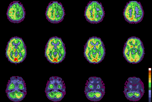
F-18 FDG PET axial images
View main image(pt) in a separate image viewer
Full history/Diagnosis is available below
Diagnosis: Crossed cerebellar diaschisis.
Full history:
30-year old woman with history
of recurrent anaplastic ganglioglioma status post
surgery and intracavitary radiotherapy as well as
external beam radiotherapy. The magnetic resonance
imaging examination has been stable over the past
year, but her clinical symptoms of right-sided
weakness, memory loss, and emotional lability are
worsening. This examination was requested to
evaluate for evidence of residual tumor.
Radiopharmaceutical:
10.8 mCi F-18
fluorodeoxyglucose i.v.
Findings:
Subtle increased FDG accumulation,
less than that of normal gray matter, was seen
corresponding to the rim enhancement at the tumor
resection bed in the left parietal lobe seen on magnetic
resonance imaging (not shown). There is diffusely
decreased tracer accumulation throughout the left
cerebral hemisphere, predominantly involving the
frontal, parietal, and temporal lobes. Decreased
activity also is seen in the left basal ganglia and
thalamus. Decreased tracer accumulation is seen in
the right cerebellar hemisphere, likely reflecting
crossed cerebellar diaschisis. This diffusely decreased
metabolism in the territories above is new since the
patientıs prior PET imaging study performed 17
months ago. These findings are thought to be most
consistent with the effects of radiation on the cerebral
vessels and blood flow.
Discussion:
Crossed cerebellar diaschisis
refers to hypometabolism in a cerebellar hemisphere
contralateral to a cerebral hemispheric lesion. The
location of the lesion is a critical factor in producing
this effect as lesions located in the motor cortex,
anterior corona radiata, and thalamus produce the
most marked suppression of the contralateral
cerebellar cortical metabolism. The cerebellar
hemispheric hypometabolism is thought to be
secondary to disconnection of cerebro-ponto-cerebellar
pathways, which leads to decreased oxygen and
glucose utilization and, hence, decreased CO2
production, which results in local arterial constriction
(decreased cerebellar blood flow) and diminished
uptake of tracer. The causes of crossed cerebellar
diaschisis include stroke, brain tumor, and sickle cell
disease. The ipsilateral hypometabolism in the
pontine, thalamic, and basal ganglia in this patient
was thought to be due to the known interval
radiotherapy (stereotactic radiosurgery 11 months
prior to the current study), although the presence of
residual tumor also raises the possibility of tumor
induced hypometabolism.
References:
1) Rumbaugh C, et al. Cerebrovascular disease:
imaging and intervention. 1995
2) Rozental, et al. Cerebral diaschisis in patients
with malignant glioma. J Neuro Oncol 1990; 8:153-
161
ACR Codes and Keywords:
References and General Discussion of PET Tumor Imaging Studies (Anatomic field:Skull and Contents, Category:Organ specific)
Search for similar cases.
Edit this case
Add comments about this case
Read comments about this case
Return to the Teaching File home page.
Case number: pt006
Copyright by Wash U MO

